News

Publication Placeholder

Lemke, S.B., and Schnorrer, F.
Mech Dev, 2016,  [Epub ahead of print].

Mechanical forces during muscle development.


Muscles are the major force producing tissue in the human body. While certain muscle types specialise in producing maximum forces, others are very enduring. An extreme example is the heart, which continuously beats for the entire life. Despite being specialised, all body muscles share similar contractile mini-machines called sarcomeres that are organised into regular higher order structures called myofibrils. The major sarcomeric components and their organisational principles are conserved throughout most of the animal kingdom. In this review, we discuss recent progress in the understanding of myofibril and sarcomere development largely obtained from in vivo models. We focus on the role of mechanical forces during muscle and myofibril development and propose a tension driven self-organisation mechanism for myofibril formation. We discuss recent technological advances that allow quantification of forces across tissues or molecules in vitro and in vivo. Although their application towards muscle development is still in its infancy, these technologies are likely to provide fundamental new insights into the mechanobiology of muscle and myofibril development in the near future.


The immune system can fight specifically against cancer by tumor-specific T cells although suitable altered target structures are currently mostly unknown. A team at the the Max Planck Institute of Biochemistry and Technical University of Munich (TUM) has developed a method that allows for the first time the reliable identification of suitable antigens directly from patients` tumor cells by mass spectrometry. These structures proved to be immunogenic. This procedure therefore opens up new possibilities for individualized targeted cancer treatments.

Through evolution, the immune system has developed sophisticated mechanisms for fighting illnesses associated with viruses and tumors. T cells play an important role in this setting. They can identify peptides, small protein sturctures, presented by the body`s own cells that may indicate a viral infection or a genetic alteration as for example a mutation – a characteristic of tumor cells. Peptides identified by immune cells are known as antigens. T cells that recognize antigens can trigger a reaction that destroys the targeted cells. In recent years research teams, including TUM researchers, have successfully utilized this characteristic for cancer treatments. Different approaches have emerged. Vaccinating a patient with an antigen can stimulate the body to enhance the production of specific T cells. Another possibility is to enrich T cells that are "trained" for certain antigens and transfer them to the patient.

A method, that identifies the antigen structures of patient specific tumors has now been developed by a team led by Angela M. Krackhardt, a professor of translational immunotherapy at the Third Medical Clinic at TUM's Klinikum rechts der Isar, and Professor Matthias Mann of the Department of Proteomics and Signal Transduction at the Max Planck Institute of Biochemistry. Krackhardt and Mann have described their approach in an article published in the Journal Nature Communications. Unlike former applied methods, it is not based on predictive models. Instead, a mass spectrometer is used to identify the peptides actually present on the tumor surface.

read more


Publication Placeholder

Falkner, S., Grade, S., Dimou, L., Conzelmann, K.K., Bonhoeffer, T., Gotz, M., and Hubener, M.
Nature, 2016, 539, 248-253.

Transplanted embryonic neurons integrate into adult neocortical circuits.

The ability of the adult mammalian brain to compensate for neuronal loss caused by injury or disease is very limited. Transplantation aims to replace lost neurons, but the extent to which new neurons can integrate into existing circuits is unknown. Here, using chronic in vivo two-photon imaging, we show that embryonic neurons transplanted into the visual cortex of adult mice mature into bona fide pyramidal cells with selective pruning of basal dendrites, achieving adult-like densities of dendritic spines and axonal boutons within 4-8 weeks. Monosynaptic tracing experiments reveal that grafted neurons receive area-specific, afferent inputs matching those of pyramidal neurons in the normal visual cortex, including topographically organized geniculo-cortical connections. Furthermore, stimulus-selective responses refine over the course of many weeks and finally become indistinguishable from those of host neurons. Thus, grafted neurons can integrate with great specificity into neocortical circuits that normally never incorporate new neurons in the adult brain.


When it comes to recovering from insult, the adult human brain has very little ability to compensate for nerve-cell loss. Biomedical researchers and clinicians are therefore exploring the possibility of using transplanted nerve cells to replace neurons that have been irreparably damaged as a result of trauma or disease. However, it is not clear whether transplanted neurons can be integrated sufficiently, to result in restored function of the lesioned network. Now researchers at the Max Planck Institute of Neurobiology in Martinsried, the Ludwig Maximilians University Munich and the Helmholtz Zentrum München have demonstrated that, in mice, transplanted embryonic nerve cells can indeed be incorporated into an existing network and correctly carry out the tasks of damaged cells originally found in that region.

Neurodegenerative diseases such as Alzheimer's or Parkinson's disease, but also stroke or certain injuries lead to a loss of brain cells. The mammalian brain can replace these cells only in very limited areas, making the loss in most cases a permanent one. The transplantation of young nerve cells into an affected network of patients for example with Parkinson's disease, allow for the possibility of a medical improvement of clinical symptoms. However, if the nerve cells transplanted in such studies help to overcome existing network gaps or whether they actually replace the lost cells, remained unknown.

In the joint study, researchers of the Max Planck Institute of Neurobiology, the Ludwig Maximilians University Munich and the Helmholtz Zentrum München have specifically asked whether transplanted embryonic nerve cells can functionally integrate into the visual cortex of adult mice. The study was supported by the center grant (SFB) 870 of the German Research Foundation (DFG). “This brain region is ideal for such experiments,” says Magdalena Götz, joint leader of the study together with Mark Hübener, who continues to explain: “By now, we know so much about the functions of the nerve cells in the visual cortex and the connections between them that we can readily assess whether the new nerve cells actually perform the tasks normally carried out by the network.”

read more


Publication Placeholder

Park, D.I., Dournes, C., Sillaber, I., Uhr, M., Asara, J.M., Gassen, N.C., Rein, T., Ising, M., Webhofer, C., Filiou, M.D., Muller, M.B., and Turck, C.W.
Sci Rep, 2016, 6 , 35317.

Purine and pyrimidine metabolism: Convergent evidence on chronic antidepressant treatment response in mice and humans.

Selective Serotonin Reuptake Inhibitors (SSRIs) are commonly used drugs for the treatment of psychiatric diseases including major depressive disorder (MDD). For unknown reasons a substantial number of patients do not show any improvement during or after SSRI treatment. We treated DBA/2J mice for 28 days with paroxetine and assessed their behavioral response with the forced swim test (FST). Paroxetine-treated long-time floating (PLF) and paroxetine-treated short-time floating (PSF) groups were stratified as proxies for drug non-responder and responder mice, respectively. Proteomics and metabolomics profiles of PLF and PSF groups were acquired for the hippocampus and plasma to identify molecular pathways and biosignatures that stratify paroxetine-treated mouse sub-groups. The critical role of purine and pyrimidine metabolisms for chronic paroxetine treatment response in the mouse was further corroborated by pathway protein expression differences in both mice and patients that underwent chronic antidepressant treatment. The integrated -omics data indicate purine and pyrimidine metabolism pathway activity differences between PLF and PSF mice. Furthermore, the pathway protein levels in peripheral specimens strongly correlated with the antidepressant treatment response in patients. Our results suggest that chronic SSRI treatment differentially affects purine and pyrimidine metabolisms, which may explain the heterogeneous antidepressant treatment response and represents a potential biosignature.


Before a cell divides, it must first handle a large-scale project: Its entire genetic material has to be duplicated so that each of the two daughter cells is equipped with a full copy after cell division. As errors in this DNA replication could lead to the death of the cell, the process is rigorously controlled. It takes place in two phases. Researchers at the Max Planck Institute of Biochemistry in Martinsried have now revealed in the journal Cell Reports that these two phases are strictly separated from one another by breaks, thereby preventing errors in the DNA replication.

Elbphilharmonie, Berlin Airport, Stuttgart 21 – large-scale projects are frequently susceptible to errors and these are generally very cost-intensive. A cell’s most important large-scale project is the replication of its DNA, i.e. the complete duplication of its genetic material. Here, errors such as the inadvertent multiplication of a DNA sequence can change the structure of the chromosomes. This can lead to cell death or, in the case of multi-cellular organisms such as humans, to the development of cancer. For this highly important project, the cell increases its success rate by dividing the process of DNA replication into a planning phase, known as the “licensing” phase, and an implementation phase, known as the “firing” phase. The two phases follow in sequence. Boris Pfander, head of the “DNA Replication and Genome Integrity” research group, and his team have demonstrated that the baker’s yeast S. cerevisiae separates the timing of these phases from one another and that the Sld2 protein plays an important role in this regulation. “A crucial factor in the success of the DNA duplication project is, on the one hand, that project planning is completed before the building work begins, but also that no new plans are made while the actual building work is being carried out,” Pfander explains.

read more...


The so-called Holliday structure has nothing to do with holidays or vacation, but is rather a term that describes a complex crossover of similar DNA strings. Researcher Robin Holliday proposed this structure of genetic information in the 1960s. It occurs in DNA repair processes in our cells or during the formation of egg and sperm cells when paternal and maternal genetic information are combined. As reported by the journal eLife, scientists from the Max Planck Institute of Biochemistry in Martinsried have decoded the structure of the GEN1 protein that can undo this crossover, much like a molecular master tailor, by cutting the DNA with the highest precision in the correct place.

Our genetic information is stored in every cell of the body in a DNA double-strand molecule. These can be damaged by chemicals and ultraviolet light, which can in turn lead to breakages in the DNA strings. Many different repair mechanisms have been developed over the course of evolution to repair DNA damage and thereby ensure the survival of various organisms.

To understand the individual repair steps in the cells, Christian Biertümpfel, who leads the Molecular Mechanisms of DNA Repair Research Group, together with his team examined the molecular structure of the GEN1 protein. GEN1 plays an important role in the repair of DNA double-strand breaks. The repair involves a complex crossover of very similar DNA sections, which enables the faulty area to be repaired. The Holliday structure that emerges is then resolved again by GEN1 at the end of the repair process.

“With the aid of X-ray diffraction analysis, we were able to look at the positions of the individual amino acids, which are the basic building blocks of proteins. This allowed us to draw conclusions as to the exact structure and function of GEN1. We can now show that GEN1 works much like a molecular master tailor, inserting precisely symmetric cuts into the Holliday structure of the DNA”, explains Biertümpfel. “What is particularly notable is that GEN1 has a so-called chromodomain, which acts like a tailor’s straightedge. The chromodomain brings the DNA to be cut into the optimum position and increases the efficiency of GEN1”.

read more


Tobias Bonhoeffer appointed advisor of Chan Zuckerberg Science

In December 2015, Facebook founder Mark Zuckerberg and his wife Priscilla Chan announced their plan to put 99% percent of their Facebook shares, worth about 45 billion dollars, into a new project focusing on the improvement of human potential and the promotion of equality. After an initial focus on education, the initiative has now announced its second focal point: the promotion of basic science research with the goal of curing, preventing or managing all disease by the end of this century. This new project, led by neuroscientist Cori Bargmann, was announced in San Francisco yesterday. In addition to the President, the two founders also introduced the scientists who will help build this initiative in the future. The panel of experts also includes one German researcher: Tobias Bonhoeffer, Director at the Max Planck Institute of Neubiology.

Shortly after the birth of their daughter Maxima Chan Zuckerberg, the couple set up the Chan Zuckerberg Initiative, stating they wished to see their daughter grow up in a world in which it was possible to cure diseases, personalize learning, and connect people. At a press conference on the Mission Bay campus of the University of California in San Francisco yesterday, Chan and Zuckerberg announced the second cornerstone of the initiative: 'Chan Zuckerberg Science'.

Over the past few months, the paediatrician Chan and Facebook founder Zuckerberg had met with numerous scientists, engineers, and other experts. This convinced them of the importance to invest into basic research to achieve much needed scientific progress essential for combating diseases. As a result, they decided to set up "Chan Zuckerberg Science (CZS)". The work of the initiative will have three core elements: More and better cooperation of scientists and engineers, the development of new methods and technologies, and educating the general public about the benefits of scientific discovery with the result of a much more general support and willingness to donate to science.

The only European member of the Scientific Advisory Board is Max Planck Director Tobias Bonhoeffer from Martinsried. "I am truly thrilled to be part of this exciting initiative. I am convinced that CZS can provide a powerful new impetus for imaginative and forward-looking science in the biomedical arena", was his first reaction. "It is a great pleasure and honor for me to have been appointed as advisor," said Bonhoeffer, who had been in contact with Chan and Zuckerberg since their visit to Berlin earlier this year. Being involved in major science policy decisions is however nothing new for the 56-year-old neurobiologist Tobias Bonhoeffer. In his capacity as chairman of the Biology and Medicine Section of the Max Planck Society (2008 - 2011) and as a Governor of the Wellcome Trust, one of the world's largest charities, he has provided guidance and expertise to many decision-making bodies.

Read more


Changes in gene activity are fundamental to many neuropsychiatric disorders. However, the nature of the genes affected, the time and place in which these changes occur, or the impact of these changes on nerve cells, remain largely unknown. One of the reasons for this ignorance is the inability to tag individual genes and their products without disrupting normal cell functions. An international team from the Max Planck Institute of Neurobiology, the University of Birmingham, the Wigner Institute in Budapest and Femtonics Ltd. now aim to close this gap in methodology. The European Union supports the project with a Horizon 2020 grant, worth more than four million Euros.

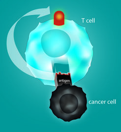
Many diseases also have a genetic component. For example, gene mutations can lead to the development of cancer or psychiatric disorders such as schizophrenia, depression or autism. Most of these diseases seem to be caused not by a single mutation but are rather the result of the interplay of dozens, maybe even hundreds of altered gene functions. It would therefore be of great benefit for the investigation, diagnosis and therapy of these illnesses, to identify the affected genes and monitor their activity in the brain of the living organism.

An interdisciplinary team around Herwig Baier from the Max Planck Institute of Neurobiology, Attila Sik from the University of Birmingham, Miklós Veres from the Wigner Institute and the microscope developers at Femtonics Ltd. aim to develop such a method. (more)


Ephrins (blue) 
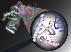
and Ephs (red) form complexes (yellow) at cell contact points.

When cells grow and divide, they come into contact with other cells. This happens not only during development and regeneration and after injury, but also during cancer growth and the formation of metastases. When cells come into contact with each other in this way, information is exchanged by proteins, which are embedded in the cell membranes and form tight lock-and-key complexes with each other. These connections must be severed if the cells want to transmit a repulsion signal. It appears that the fastest way to do this is for the cells to engulf the protein complex from the membrane of the neighbouring cell. Scientists from the Max Planck Institute of Neurobiology in Martinsried have now identified the molecules that control this process.

Development is an extremely rapid process. Increasing numbers of cells are formed which must find their correct position in the body, clearly demarcate themselves from each other to form tissue, or – as is the case in the nervous system – establish contact with partner cells in remote locations. “The crowding is accompanied by orderly pushing and shoving,” says Rüdiger Klein, whose Department at the Max Planck Institute of Neurobiology studies how cells get their bearings. “A popular way for one cell to show another which direction to take is for it to repel the other cell following brief contact.” According to the scientists’ observations, the cells do not exactly treat each other with kid gloves and even go so far as to engulf entire pieces from the membranes of other cells. More