News

Publication Placeholder

Vincenz-Donnelly, L., Holthusen, H., Korner, R., Hansen, E.C., Presto, J., Johansson, J., Sawarkar, R., Hartl, F.U., and Hipp, M.S.
EMBO J, 2017, [Epub ahead of print].
doi 10.15252/embj.201695841

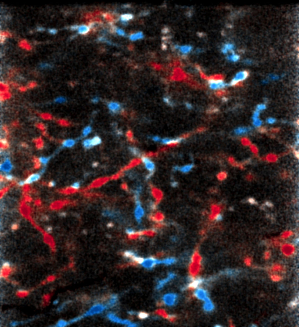
High capacity of the endoplasmic reticulum to prevent secretion and aggregation of amyloidogenic proteins

Protein aggregation is associated with neurodegeneration and various other pathologies. How specific cellular environments modulate the aggregation of disease proteins is not well understood. Here, we investigated how the endoplasmic reticulum (ER) quality control system handles β-sheet proteins that were designed de novo to form amyloid-like fibrils. While these proteins undergo toxic aggregation in the cytosol, we find that targeting them to the ER (ER-β) strongly reduces their toxicity. ER-β is retained within the ER in a soluble, polymeric state, despite reaching very high concentrations exceeding those of ER-resident molecular chaperones. ER-β is not removed by ER-associated degradation (ERAD) but interferes with ERAD of other proteins. These findings demonstrate a remarkable capacity of the ER to prevent the formation of insoluble β-aggregates and the secretion of potentially toxic protein species. Our results also suggest a generic mechanism by which proteins with exposed β-sheet structure in the ER interfere with proteostasis.


Publication Placeholder

Albert, S., Schaffer, M., Beck, F., Mosalaganti, S., Asano, S., Thomas, H.F., Plitzko, J.M., Beck, M., Baumeister, W., and Engel, B.D.
Proc Natl Acad Sci USA, 2017, [Epub ahead of print].
doi: 10.1073/pnas.1716305114

Proteasomes tether to two distinct sites at the nuclear pore complex.

The partitioning of cellular components between the nucleus and cytoplasm is the defining feature of eukaryotic life. The nuclear pore complex (NPC) selectively gates the transport of macromolecules between these compartments, but it is unknown whether surveillance mechanisms exist to reinforce this function. By leveraging in situ cryo-electron tomography to image the native cellular environment of Chlamydomonas reinhardtii, we observed that nuclear 26S proteasomes crowd around NPCs. Through a combination of subtomogram averaging and nanometer-precision localization, we identified two classes of proteasomes tethered via their Rpn9 subunits to two specific NPC locations: binding sites on the NPC basket that reflect its eightfold symmetry and more abundant binding sites at the inner nuclear membrane that encircle the NPC. These basket-tethered and membrane-tethered proteasomes, which have similar substrate-processing state frequencies as proteasomes elsewhere in the cell, are ideally positioned to regulate transcription and perform quality control of both soluble and membrane proteins transiting the NPC.


Publication Placeholder

Jaepel, J., Hubener, M., Bonhoeffer, T., and Rose, T.
Nat Neurosci, 2017, 20, 1708-1714.
doi: 10.1038/s41593-017-0021-0

Lateral geniculate neurons projecting to primary visual cortex show ocular dominance plasticity in adult mice.

Experience-dependent plasticity in the mature visual system is widely considered to be cortical. Using chronic two-photon Ca2+ imaging of thalamic afferents in layer 1 of binocular visual cortex, we provide evidence against this tenet: the respective dorsal lateral geniculate nucleus (dLGN) cells showed pronounced ocular dominance (OD) shifts after monocular deprivation in adult mice. Most (86%), but not all, of dLGN cell boutons were monocular during normal visual experience. Following deprivation, initially deprived-eye-dominated boutons reduced or lost their visual responsiveness to that eye and frequently became responsive to the non-deprived eye. This cannot be explained by eye-specific cortical changes propagating to dLGN via cortico-thalamic feedback because the shift in dLGN responses was largely resistant to cortical inactivation using the GABAA receptor agonist muscimol. Our data suggest that OD shifts observed in the binocular visual cortex of adult mice may at least partially reflect plasticity of eye-specific inputs onto dLGN neurons.


Publication Placeholder

Frauenstein, A., and Meissner, F.
Methods Mol Biol, 2018, 1714, 215-227.
doi: 10.1007/978-1-4939-7519-8_14

Quantitative Proteomics of Secreted Proteins.

Secreted proteins such as cytokines, interleukins, growth factors, and hormones have pleiotropic functions and facilitate intercellular communication in organisms. Quantification of these proteins conventionally relies on antibody-based methods, i.e., enzyme-linked immunosorbent assays (ELISA), whose large-scale use is limited by availability, specificity, and affordability.Here, we describe an experimental and bioinformatics workflow to comprehensively quantify cellular protein secretion by mass spectrometry. Secreted proteins are collected in vitro or ex vivo, digested with proteases and the resulting peptide mixtures are analyzed in single liquid chromatography-mass spectrometry (LC-MS/MS) runs. Label-free quantification and bioinformatics analysis is conducted in the MaxQuant and Perseus computational environment. Our workflow allows the quantification of thousands of secreted proteins spanning a concentration range of four orders of magnitude and permits the systems-level characterization of secretory programs as well as the discovery of proteins with unexpected extracellular functions.


Publication Placeholder

Hosp, F.*, Gutierrez-Angel, S.*, Schaefer, M.H., Cox, J., Meissner, F., Hipp, M.S., Hartl, F.U., Klein, R., Dudanova, I., and Mann, M.
Cell Rep, 2017, 21, 2291-2303.
* equal contribution

Spatiotemporal Proteomic Profiling of Huntington's Disease Inclusions Reveals Widespread Loss of Protein Function.

Aggregation of polyglutamine-expanded huntingtin exon 1 (HttEx1) in Huntington's disease (HD) proceeds from soluble oligomers to late-stage inclusions. The nature of the aggregates and how they lead to neuronal dysfunction is not well understood. We employed mass spectrometry (MS)-based quantitative proteomics to dissect spatiotemporal mechanisms of neurodegeneration using the R6/2 mouse model of HD. Extensive remodeling of the soluble brain proteome correlated with insoluble aggregate formation during disease progression. In-depth and quantitative characterization of the aggregates uncovered an unprecedented complexity of several hundred proteins. Sequestration to aggregates depended on protein expression levels and sequence features such as low-complexity regions or coiled-coil domains. In a cell-based HD model, overexpression of a subset of the sequestered proteins in most cases rescued viability and reduced aggregate size. Our spatiotemporally resolved proteome resource of HD progression indicates that widespread loss of cellular protein function contributes to aggregate-mediated toxicity.


The cerebral cortex is where we learn and think, form impressions of our environment, control conscious behaviour, and store memories. According to the textbooks, the upstream regions of the brain like the thalamus only contribute to these processes by forwarding information from the sensory organs to the corresponding regions of the cerebral cortex and filtering the information, if necessary. Scientists from the Max Planck Institute of Neurobiology have now shown that the textbook account will have to be revised in part. In the mouse brain, at least, the thalamus appears to play a considerably more active role in visual processing in the context of learning than was previously assumed.

A young brain has much to learn – including how it should interpret information from both eyes and collate it into a meaningful image of the environment. Hence, the cells in the visual cortex establish connections with each other during brain development to enable the optimum processing of visual environmental stimuli. In some cases, however, the signals from one eye do not correspond with those from the other eye, for example in children with strabismus. This can result in the incorrect “wiring” of the eyes to the cerebral cortex. The resulting visual weakness can often be corrected by temporarily covering the dominant eye. If this is done during the critical phase for the development of visual processing, the cells alter their connections, and the brain area responsible for the dominant eye receives signals from the uncovered, weaker eye.

The brain can thus learn to process the visual information differently – an insight that is applied successfully through the use of eye patches in children with strabismus. These well-researched processes in the visual cortex have also been used for many years as a model for the study of learning mechanisms in the cerebral cortex based on the example of the mouse brain.

When scientists from Tobias Bonhoeffer’s department examined the activity of neurons from upstream brain areas, in particular the thalamus, during a temporary closure of the eye, they made an astonishing discovery: these cells did not simply relay information from the eyes to the cerebral cortex but also altered their signals in response to the eye closure. “This was completely unexpected, as it has been believed for over 50 years that the thalamus only forwards information and is not actively involved in learning processes,” reports Tobias Rose, leader of the study.

read more


Publication Placeholder

Weckmann, K., Deery, M.J., Howard, J.A., Feret, R., Asara, J.M., Dethloff, F., Filiou, M.D., Iannace, J., Labermaier, C., Maccarrone, G., Webhofer, C., Teplytska, L., Lilley, K., Muller, M.B., and Turck, C.W.
Sci Rep, 2017, 7, 15788.

Ketamine's antidepressant effect is mediated by energy metabolism and antioxidant defense system.

Fewer than 50% of all patients with major depressive disorder (MDD) treated with currently available antidepressants (ADs) show full remission. Moreover, about one third of the patients suffering from MDD does not respond to conventional ADs and develop treatment-resistant depression (TRD). Ketamine, a non-competitive, voltage-dependent N-Methyl-D-aspartate receptor (NMDAR) antagonist, has been shown to have a rapid antidepressant effect, especially in patients suffering from TRD. Hippocampi of ketamine-treated mice were analysed by metabolome and proteome profiling to delineate ketamine treatment-affected molecular pathways and biosignatures. Our data implicate mitochondrial energy metabolism and the antioxidant defense system as downstream effectors of the ketamine response. Specifically, ketamine tended to downregulate the adenosine triphosphate (ATP)/adenosine diphosphate (ADP) metabolite ratio which strongly correlated with forced swim test (FST) floating time. Furthermore, we found increased levels of enzymes that are part of the ‘oxidative phosphorylation’ (OXPHOS) pathway. Our study also suggests that ketamine causes less protein damage by rapidly decreasing reactive oxygen species (ROS) production and lend further support to the hypothesis that mitochondria have a critical role for mediating antidepressant action including the rapid ketamine response.


Publication Placeholder

Balsevich, G., Hausl, A.S., Meyer, C.W., Karamihalev, S., Feng, X., Pohlmann, M.L., Dournes, C., Uribe-Marino, A., Santarelli, S., Labermaier, C., Hafner, K., Mao, T., Breitsamer, M., Theodoropoulou, M., Namendorf, C., Uhr, M., Paez-Pereda, M., Winter, G., Hausch, F., Chen, A., Tschop, M.H., Rein, T., Gassen, N.C., and Schmidt, M.V.
Nat Commun, 2017, 8, 1725.

Stress-responsive FKBP51 regulates AKT2-AS160 signaling and metabolic function.

The co-chaperone FKBP5 is a stress-responsive protein-regulating stress reactivity, and its genetic variants are associated with T2D related traits and other stress-related disorders. Here we show that FKBP51 plays a role in energy and glucose homeostasis. Fkbp5 knockout (51KO) mice are protected from high-fat diet-induced weight gain, show improved glucose tolerance and increased insulin signaling in skeletal muscle. Chronic treatment with a novel FKBP51 antagonist, SAFit2, recapitulates the effects of FKBP51 deletion on both body weight regulation and glucose tolerance. Using shorter SAFit2 treatment, we show that glucose tolerance improvement precedes the reduction in body weight. Mechanistically, we identify a novel association between FKBP51 and AS160, a substrate of AKT2 that is involved in glucose uptake. FKBP51 antagonism increases the phosphorylation of AS160, increases glucose transporter 4 expression at the plasma membrane, and ultimately enhances glucose uptake in skeletal myotubes. We propose FKBP51 as a mediator between stress and T2D development, and potential target for therapeutic approaches.


Publication Placeholder

Thoma V., Kobayashi K. and Tanimoto H.
eNeuro 4, 2017

The Role of the Gustatory System in the Coordination of Feeding

To survive, all animals must find, inspect and ingest food. Behavioral coordination and control of feeding is therefore a challenge that animals must face. Here, we focus on how the gustatory system guides the precise execution of behavioral sequences that promote ingestion and suppresses competing behaviors. We summarize principles learnt from Drosophila, where underlying sensory neuronal mechanisms are illustrated in great detail. Moreover, we compare these principles with findings in other animals, where such coordination plays prominent roles. These examples suggest that the use of gustatory information for feeding coordination has an ancient origin and is prevalent throughout the animal kingdom.


Atlas of the Heart - A healthy heart beats about two billion times during a lifetime – thanks to the interplay of more than 10,000 proteins. Researcher from the Max Planck Institute of Biochemistry (MPIB) and the German Heart Centre at the Technical University of Munich (TUM) have now determined which and how many individual proteins are present in each type of cell that occurs in the heart. In doing so, they compiled the first atlas of the healthy human heart, known as the cardiac proteome. The atlas will make it easier to identify differences between healthy and diseased hearts in future.

Proteins are the molecular machines of cells, in which they perform a range of functions. They are produced by the cells based on blueprints stored in their DNA. Changes occurring at the DNA or protein level can lead to disorders. For such changes to be recognized as underlying causes of heart disease, it is important to know precisely which proteins are present in the healthy heart and in what quantities.

Protein map of the heart
The first such protein atlas of the heart was recently published in Nature Communications by a research team from Munich. The scientists determined the protein profile of cells in all the regions of the heart, such as heart valves, cardiac chambers and major blood vessels. In addition, they investigated the protein composition in three different cell types of the heart: the cardiac fibroblasts, the smooth muscle cells and the endothelial cells. In this way the researchers were able to map the distribution of proteins in the various regions of the heart. Using mass spectrometry, they identified nearly 11,000 different proteins throughout the heart.

Previous studies had focussed for the most part only on individual cell types, or they used tissue from diseased hearts. "This approach has two problems," says Sophia Doll of the MPIB and lead author of the study. "First, the results did not give a full picture of the heart across all its regions and tissues; and second, comparative data on healthy hearts were often missing. Our study has eliminated both problems. Now the data can be used as a reference for future studies."

"Looking at the protein atlas of the human heart, you can see that all healthy hearts work in a very similar manner. We measured similar protein compositions in all the regions with few differences between them," says Sophia Doll. We were also surprised to find that the right and left halves of the heart are similar, despite having quite different functions: the right half pumps oxygen-poor blood to the lungs, while the left half pumps oxygen-rich blood from the lungs to the body.

read more