News

Publication Placeholder

Monkemeyer, L., Klaips, C.L., Balchin, D., Korner, R., Hartl, F.U., and Bracher, A.
Mol Cell, 2019, [Epub ahead of print].
doi: 10.1016/j.molcel.2019.01.034

Chaperone Function of Hgh1 in the Biogenesis of Eukaryotic Elongation Factor 2

Eukaryotic elongation factor 2 (eEF2) is an abundant and essential component of the translation machinery. The biogenesis of this 93 kDa multi-domain protein is assisted by the chaperonin TRiC/CCT. Here, we show in yeast cells that the highly conserved protein Hgh1 (FAM203 in humans) is a chaperone that cooperates with TRiC in eEF2 folding. In the absence of Hgh1, a substantial fraction of newly synthesized eEF2 is degraded or aggregates. We solved the crystal structure of Hgh1 and analyzed the interaction of wild-type and mutant Hgh1 with eEF2. These experiments revealed that Hgh1 is an armadillo repeat protein that binds to the dynamic central domain III of eEF2 via a bipartite interface. Hgh1 binding recruits TRiC to the C-terminal eEF2 module and prevents unproductive interactions of domain III, allowing efficient folding of the N-terminal GTPase module. eEF2 folding is completed upon dissociation of TRiC and Hgh1.


Publication Placeholder

Cost, A.L., Khalaji, S., and Grashoff, C.
Curr Protoc Cell Biol, 2019, e85, [Epub ahead of print].
doi: 10.1002/cpcb.85

Genetically Encoded FRET-Based Tension Sensors

Genetically encoded Förster resonance energy transfer (FRET)-based tension sensors measure piconewton-scale forces across individual molecules in living cells or whole organisms. These biosensors show comparably high FRET efficiencies in the absence of tension, but FRET quickly decreases when forces are applied. In this article, we describe how such biosensors can be generated for a specific protein of interest, and we discuss controls to confirm that the observed differences in FRET efficiency reflect changes in molecular tension. These FRET efficiency changes can be related to mechanical forces as the FRET-force relationship of the employed tension sensor modules are calibrated. We provide information on construct generation, expression in cells, and image acquisition using live-cell fluorescence lifetime imaging microscopy (FLIM). Moreover, we describe how to analyze, statistically evaluate, and interpret the resulting data sets. Together, these protocols should enable the reader to plan, execute, and interpret FRET-based tension sensor experiments.


Much insight into the brain’s function has been gained from scientists working with animal models. However, animal models have their limitations since animal brains undergo a very different development process to humans brains. Some research has been carried out on the human brain, but it has relied on post-mortem donations and cultured cells. Since human brain tissue is difficult to obtain and animal models can only model the human brain to a limited extent, there was a crucial gap in brain research and a novel method to model human disease was highly sought after.

The Cappello research group at the Max Planck Institute of Psychiatry in a highly collaborative work, particularly with the laboratory of Barbara Treutlein from the Max Planck Institute for Evolutionary Anthropology in Leipzig, have employed brain organoids to model developmental brain malfunctions. These brain organoids have revolutionized neuroscience as they can recapitulate the way neurons differentiate in development to a remarkably high degree. When the human brain develops, new cells grow and aggregate in a very specific order.

The brain organoids in this study were grown from skin cell biopsies donated by patients. Silvia Cappello, who leads the work, explains: “We reprogrammed the skin cells into induced pluripotent stem cells. These pluripotent stem cells were then used to generate brain cells, which could be differentiated into many different types of brain cells.” The different types of brain cells and their interactions can then be studied behaving in a way they would in an actual human brain. Cappello adds: “Brain organoids give us a much clearer picture of how brain cells are functioning and can accurately model human neurological diseases.”

The ability to model human brain development in vitro holds tremendous translational value. Brain organoids have already helped scientists to better understand the Zika virus, Alzheimer’s disease and autism. As the brain organoid is grown from the cells of an individual, it allows scientists to study exactly what is happening in individual patients. Cappello concludes: “Validating brain organoids represents a hugely important step in helping us to understand developmental and neurological disorders and they hold great promise for discovering new treatments.”

read more


Publication Placeholder

Schopf, F.H., Huber, E.M., Dodt, C., Lopez, A., Biebl, M.M., Rutz, D.A., Muhlhofer, M., Richter, G., Madl, T., Sattler, M., Groll, M., and Buchner, J.
Mol Cell, 2019, [Epub ahead of print].
doi: 10.1016/j.molcel.2019.02.011

The Co-chaperone Cns1 and the Recruiter Protein Hgh1 Link Hsp90 to Translation Elongation via Chaperoning Elongation Factor 2

The Hsp90 chaperone machinery in eukaryotes comprises a number of distinct accessory factors. Cns1 is one of the few essential co-chaperones in yeast, but its structure and function remained unknown. Here, we report the X-ray structure of the Cns1 fold and NMR studies on the partly disordered, essential segment of the protein. We demonstrate that Cns1 is important for maintaining translation elongation, specifically chaperoning the elongation factor eEF2. In this context, Cns1 interacts with the novel co-factor Hgh1 and forms a quaternary complex together with eEF2 and Hsp90. The in vivo folding and solubility of eEF2 depend on the presence of these proteins. Chaperoning of eEF2 by Cns1 is essential for yeast viability and requires a defined subset of the Hsp90 machinery as well as the identified eEF2 recruiting factor Hgh1.


Our world is full of sensory stimuli. Depending on what we see, smell, taste, feel, or hear, we are compelled to behave in a predictable way – like approaching tasty food or avoiding an oncoming car. The brain’s ability to make sense of the diverse sensory stimuli and to coordinate the appropriate behavioral response relies critically on the function of the cerebellum. This hindbrain region, critical to sensorimotor coordination, is conserved across vertebrates, from humans to birds to fish.

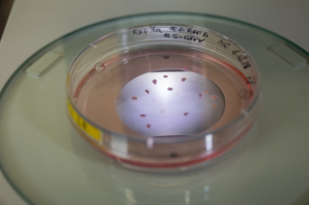
The mammalian cerebellum, however, contains hundreds of thousands of Purkinje cells, each receiving inputs from many thousands of presynaptic neurons. Cracking the cerebellar code here is nearly impossible, even with the latest methods. Ruben Portugues and his team thus focus on a "simpler" version: the cerebellum of six to eight day old zebrafish larvae.

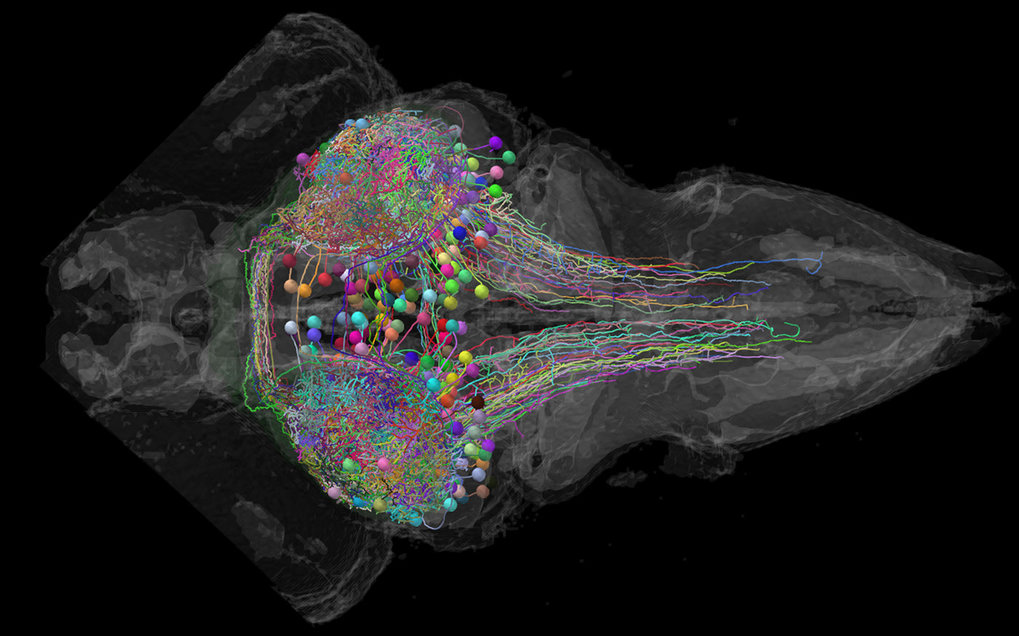
“At this age, the zebrafish cerebellum contains about 500 Purkinje cells and is involved in behaviors such as swimming and eye movements”, explains Laura Knogler, who studied the cerebellar circuits together with graduate student Andreas Kist. “It’s all there and still very complex, but we have a chance to see all cells’ activity in the transparent brains of these fish and directly record the detailed activity of individual cells.” By studying the cerebellum of zebrafish larvae behaving within virtual-reality environments, the scientists were now able to tackle the central question of how the cerebellum coordinates behavior.

Like many vertebrates, zebrafish use visual cues to direct their movements, keep track of their environment or to identify potential predators or prey. Using this knowledge, the neuroscientists showed the fish different visual stimuli while observing neuronal activity and the motor functions. The surprising result was a cerebellar division into three behavioral modules, each encoding a distinct type of visual information: directional motion onset, rotational motion velocity, or changes in luminance. Every studied Purkinje cell belonged to one of these three modules.

In contrast, the behavior of the fish was encoded in nearly the same way by all cells. This became visible in an impressive way when the fish were swimming: “Nearly the entire cerebellum lit up with fluorescence, showing an overwhelming amount of Purkinje cell excitation during swim bouts”, relates Andreas Kist. The scientists believe that the observed cerebellar organization is an important trait for neural coding and associative learning: “The modules appeared optimized to organize the information necessary for the principal behaviors of the zebrafish at this age, yet may also allow for the flexibility required to learn new things through experience”, explains Knogler. “I wouldn’t be surprised if other sensory input and the cerebella of other species are organized in a similar way.”.

read more


Publication Placeholder

Bantele, S.C.S., Lisby, M., and Pfander, B.
Nat Commun, 2019, 10, 944.
doi: 10.1038/s41467-019-08889-5

Quantitative sensing and signalling of single-stranded DNA during the DNA damage response

The DNA damage checkpoint senses the presence of DNA lesions and controls the cellular response thereto. A crucial DNA damage signal is single-stranded DNA (ssDNA), which is frequently found at sites of DNA damage and recruits the sensor checkpoint kinase Mec1-Ddc2. However, how this signal - and therefore the cell's DNA damage load - is quantified, is poorly understood. Here, we use genetic manipulation of DNA end resection to induce quantitatively different ssDNA signals at a site-specific double strand break in budding yeast and identify two distinct signalling circuits within the checkpoint. The local checkpoint signalling circuit leading to γH2A phosphorylation is unresponsive to increased amounts of ssDNA, while the global checkpoint signalling circuit, which triggers Rad53 activation, integrates the ssDNA signal quantitatively. The global checkpoint signal critically depends on the 9-1-1 and its downstream acting signalling axis, suggesting that ssDNA quantification depends on at least two sensor complexes.


One in four people in Western and Asian societies develop a build-up of fat in the liver as a result of an unhealthy diet. This disease – referred to as non-alcoholic fatty liver disease (NAFLD) – causes no symptoms initially but can develop into end-stage liver cirrhosis with limited treatment options. A discovery, published today in Molecular Systems Biology, paves the way for a simple blood test to detect early stages of NAFLD, opening up the possibility of preventing the development of liver cirrhosis through lifestyle changes or pharmaceutical intervention.

The liver is an important organ, filtering toxic substances from the body and producing proteins required for digestion, blood clotting, and other important physiological functions. “The liver is very resilient and capable of regenerating itself, which may be the reason why liver damages due to excessive fat deposition can go undetected for a long time,” says EMBO Member Matthias Mann of the Max Planck Institute of Biochemistry in Martinsried, Germany, and the University of Copenhagen, Denmark, who led the study. However, when damage accumulates liver function eventually starts to fail.

To date, the standard procedure for diagnosing NAFLD is liver biopsy – a cumbersome and costly procedure that can lead to complications. Non-invasive methods that reliably detect early stage NAFLD are therefore urgently required.

Matthias Mann and his colleagues investigated the plasma proteome – the entire set of proteins present in the blood plasma – of NAFLD patients. Using sophisticated mass spectrometry technologies, they uncovered a set of proteins that accumulate in the plasma of patients with non-symptomatic NAFLD.

read more


Publication Placeholder

Papadopoulou, A.A., Muller, S.A., Mentrup, T., Shmueli, M.D., Niemeyer, J., Haug-Kroper, M., von Blume, J., Mayerhofer, A., Feederle, R., Schroder, B., Lichtenthaler, S.F., and Fluhrer, R.
EMBO Rep, 2019, [Epub ahead of print].
doi: 10.15252/embr.201846451

Signal Peptide Peptidase-Like 2c (SPPL2c) impairs vesicular transport and cleavage of SNARE proteins

Members of the GxGD-type intramembrane aspartyl proteases have emerged as key players not only in fundamental cellular processes such as B-cell development or protein glycosylation, but also in development of pathologies, such as Alzheimer's disease or hepatitis virus infections. However, one member of this protease family, signal peptide peptidase-like 2c (SPPL2c), remains orphan and its capability of proteolysis as well as its physiological function is still enigmatic. Here, we demonstrate that SPPL2c is catalytically active and identify a variety of SPPL2c candidate substrates using proteomics. The majority of the SPPL2c candidate substrates cluster to the biological process of vesicular trafficking. Analysis of selected SNARE proteins reveals proteolytic processing by SPPL2c that impairs vesicular transport and causes retention of cargo proteins in the endoplasmic reticulum. As a consequence, the integrity of subcellular compartments, in particular the Golgi, is disturbed. Together with a strikingly high physiological SPPL2c expression in testis, our data suggest involvement of SPPL2c in acrosome formation during spermatogenesis.


Publication Placeholder

Cosolo, A., Jaiswal, J., Csordas, G., Grass, I., Uhlirova, M., and Classen, A.K.
Elife 8, 2019.
doi: 10.7554/eLife.41036

JNK-dependent cell cycle stalling in G2 promotes survival and senescence-like phenotypes in tissue stress

The restoration of homeostasis after tissue damage relies on proper spatial-temporal control of damage-induced apoptosis and compensatory proliferation. In Drosophila imaginal discs these processes are coordinated by the stress response pathway JNK. We demonstrate that JNK signaling induces a dose-dependent extension of G2 in tissue damage and tumors, resulting in either transient stalling or a prolonged but reversible cell cycle arrest. G2-stalling is mediated by downregulation of the G2/M-specific phosphatase String(Stg)/Cdc25. Ectopic expression of stg is sufficient to suppress G2-stalling and reveals roles for stalling in survival, proliferation and paracrine signaling. G2-stalling protects cells from JNK-induced apoptosis, but under chronic conditions, reduces proliferative potential of JNK-signaling cells while promoting non-autonomous proliferation. Thus, transient cell cycle stalling in G2 has key roles in wound healing but becomes detrimental upon chronic JNK overstimulation, with important implications for chronic wound healing pathologies or tumorigenic transformation.


We don’t need to think twice: if an object is approaching on a collision course, we quickly get out of its way. But if something captures our interest, we move directly towards it. Little is known about how the brain classifies visual objects as either attractive or threatening, and how this information is channeled to initiate an appropriate behavior. This gap in our knowledge is now being filled.

Zebrafish larvae are about five millimeters long and almost transparent, so that we can peek into their brain while it is engaged in a behavioral task. With the aid of newly developed optical and genetic methods, scientists are now able to observe the activity and activation sequence of individual nerve cells. Scientists are thus able to follow the transition from a visual perception to a behavior in real time under the microscope. What the neurobiologists discovered is that “predator” or “prey” categories each activate a dedicated nerve tract to steer behavior.

Previous studies had indicated that this activity originates in the tectum of the fish brain. Humans also have such a tectum, the superior colliculus, which is thought to have very similar functions. To understand what happens in the fish tectum at the cellular level, Thomas Helmbrecht from the Max Planck Institute of Neurobiology studied the reaction among young fish to virtual dots, while observing the activity of their nerve cells and manipulating them using optogenetic methods.

Depending on the size and animated movement, the dots were initially classified as prey or predator in the tectum of the zebrafish. The tectum then transmitted the decision made in each case to the hindbrain via one of two different, spatially separate pathways of nerve cells.

The neurons at the end of the signal chain initiated either an avoidance or approach movement, depending on which of the two pathways carried the information. The scientists were also able to demonstrate that the nerve cells transmitted precise data relating to the position of the potential prey via the approach pathway. The muscles can evidently be controlled by the neurons in such a way that the young fish is able to swim directly towards its prey.

At least 29 different nerve cell types in the tectum project information throughout the brain. “We now want to find out in detail how these individual cell types contribute to behavior,” explains Herwig Baier, in whose laboratory the experiments were conducted. “For the first time, we have the opportunity to fully reconstruct the brain activity that forms the basis of a complex behavioral decision, from the sensory input all the way to the motor output.”

read more