News

Publication Placeholder

Matscheko, N., Mayrhofer, P., Rao, Y., Beier, V., and Wollert, T.
PLoS Biol, 2019, 17, e3000377
doi: 10.1371/journal.pbio.3000377

Atg11 tethers Atg9 vesicles to initiate selective autophagy

Autophagy recycles cytoplasmic components by sequestering them in double membrane-surrounded autophagosomes. The two proteins Atg11 and Atg17 are scaffolding components of the Atg1 kinase complex. Atg17 recruits and tethers Atg9-donor vesicles, and the corresponding Atg1 kinase complex induces the formation of nonselective autophagosomes. Atg11 initiates selective autophagy and coordinates the switch to nonselective autophagy by recruiting Atg17. The molecular function of Atg11 remained, however, less well understood. Here, we demonstrate that Atg11 is activated by cargo through a direct interaction with autophagy receptors. Activated Atg11 dimerizes and tethers Atg9 vesicles, which leads to the nucleation of phagophores in direct vicinity of cargo. Starvation reciprocally regulates the activity of both tethering factors by initiating the degradation of Atg11 while Atg17 is activated. This allows Atg17 to sequester and tether Atg9 vesicles independent of cargo to nucleate nonselective phagophores. Our data reveal insights into the molecular mechanisms governing cargo selection and specificity in autophagy.


graduation

Congratulations on your PhD!

Nicole Teichmann
Preclinical evaluation of an oral MEK1/2 inhibitor as a therapeutic strategy for GEMM-based Pancreatic ductal adenocarcinoma (PDAC)
RG: Jens Siveke

Sigrun Schmähling
Biochemical purification and functional characterization of a novel trithorax-group protein complex
RG: Jürg Müller


Publication Placeholder

Cramer, K., Bolender, A.L., Stockmar, I., Jungmann, R., Kasper, R., and Shin, J.Y.
Int J Mol Sci, 2019, 20, [Epub ahead of print].
doi: 10.3390/ijms20143376

Visualization of Bacterial Protein Complexes Labeled with Fluorescent Proteins and Nanobody Binders for STED Microscopy

In situ visualization of molecular assemblies near their macromolecular scale is a powerful tool to investigate fundamental cellular processes. Super-resolution light microscopies (SRM) overcome the diffraction limit and allow researchers to investigate molecular arrangements at the nanoscale. However, in bacterial cells, visualization of these assemblies can be challenging because of their small size and the presence of the cell wall. Thus, although conceptually promising, successful application of SRM techniques requires careful optimization in labeling biochemistry, fluorescent dye choice, bacterial biology and microscopy to gain biological insights. Here, we apply Stimulated Emission Depletion (STED) microscopy to visualize cell division proteins in bacterial cells, specifically E. coli and B. subtilis. We applied nanobodies that specifically recognize fluorescent proteins, such as GFP, mCherry2 and PAmCherry, fused to targets for STED imaging and evaluated the effect of various organic fluorescent dyes on the performance of STED in bacterial cells. We expect this research to guide scientists for in situ macromolecular visualization using STED in bacterial systems.


Publication Placeholder

Kabacaoglu, D., Ruess, D.A., Ai, J., and Algul, H.
Cancers (Basel), 2019, 11.
doi: 10.3390/cancers11070937

NF-kappaB/Rel Transcription Factors in Pancreatic Cancer: Focusing on RelA, c-Rel, and RelB

Regulation of Nuclear factor kappa-light-chain-enhancer of activated B cells (NF-κB)/Rel transcription factors (TFs) is extremely cell-type-specific owing to their ability to act disparately in the context of cellular homeostasis driven by cellular fate and the microenvironment. This is also valid for tumor cells in which every single component shows heterogenic effects. Whereas many studies highlighted a per se oncogenic function for NF-κB/Rel TFs across cancers, recent advances in the field revealed their additional tumor-suppressive nature. Specifically, pancreatic ductal adenocarcinoma (PDAC), as one of the deadliest malignant diseases, shows aberrant canonical-noncanonical NF-κB signaling activity. Although decades of work suggest a prominent oncogenic activity of NF-κB signaling in PDAC, emerging evidence points to the opposite including anti-tumor effects. Considering the dual nature of NF-κB signaling and how it is closely linked to many other cancer related signaling pathways, it is essential to dissect the roles of individual Rel TFs in pancreatic carcinogenesis and tumor persistency and progression. Here, we discuss recent knowledge highlighting the role of Rel TFs RelA, RelB, and c-Rel in PDAC development and maintenance. Next to providing rationales for therapeutically harnessing Rel TF function in PDAC, we compile strategies currently in (pre-)clinical evaluation.


Publication Placeholder

Kober-Hasslacher, M., and Schmidt-Supprian, M.
Cancers (Basel), 2019, 11.
doi: 10.3390/cancers11070941

The Unsolved Puzzle of c-Rel in B Cell Lymphoma

Aberrant constitutive activation of Rel/NF-κB transcription factors is a hallmark of numerous cancers. Of the five Rel family members, c-Rel has the strongest direct links to tumorigenesis. c-Rel is the only member that can malignantly transform lymphoid cells in vitro. Furthermore, c-Rel is implicated in human B cell lymphoma through the frequent occurrence of REL gene locus gains and amplifications. In normal physiology, high c-Rel expression predominates in the hematopoietic lineage and a diverse range of stimuli can trigger enhanced expression and activation of c-Rel. Both expression and activation of c-Rel are tightly regulated on multiple levels, indicating the necessity to keep its functions under control. In this review we meta-analyze and integrate studies reporting gene locus aberrations to provide an overview on the frequency of REL gains in human B cell lymphoma subtypes, namely follicular lymphoma, diffuse large B cell lymphoma, primary mediastinal B cell lymphoma, and classical Hodgkin lymphoma. We also summarize current knowledge on c-Rel expression and protein localization in these human B cell lymphomas and discuss the co-amplification of BCL11A with REL. In addition, we highlight and illustrate key pathways of c-Rel activation and regulation with a specific focus on B cell biology.


graduation

Congratulations on your PhD!


Anna-Lena Cost
Molecular Force Measurements in Desmosomes
RG: Carsten Grashoff

Philipp Glock
Controlling and Reshaping Biological Reaction-Diffusion
RG: Petra Schwille


Publication Placeholder

Marti Fernandez, I., Macrini, C., Krumbholz, M., Hensbergen, P.J., Hipgrave Ederveen, A.L., Winklmeier, S., Vural, A., Kurne, A., Jenne, D., Kamp, F., Gerdes, L.A., Hohlfeld, R., Wuhrer, M., Kumpfel, T., and Meinl, E.
Front Immunol, 2019, 10, 1189.
doi: 10.3389/fimmu.2019.01189

The Glycosylation Site of Myelin Oligodendrocyte Glycoprotein Affects Autoantibody Recognition in a Large Proportion of Patients

Autoantibodies to myelin oligodendrocytes glycoprotein (MOG) are found in a fraction of patients with inflammatory demyelination and are detected with MOG-transfected cells. While the prototype anti-MOG mAb 8-18C5 and polyclonal anti-MOG responses from different mouse strains largely recognize the FG loop of MOG, the human anti-MOG response is more heterogeneous and human MOG-Abs recognizing different epitopes were found to be pathogenic. The aim of this study was to get further insight into details of antigen-recognition by human MOG-Abs focusing on the impact of glycosylation. MOG has one known N-glycosylation site at N31 located in the BC loop linking two beta-sheets. We compared the reactivity to wild type MOG with that toward two different mutants in which the neutral asparagine of N31 was mutated to negatively charged aspartate or to the neutral alanine. We found that around 60% of all patients (16/27) showed an altered reactivity to one or both of the mutations. We noted seven different patterns of recognition of the two glycosylation-deficient mutants by different patients. The introduced negative charge at N31 enhanced recognition in some, but reduced recognition in other patients. In 7/27 patients the neutral glycosylation-deficient mutant was recognized stronger. The folding of the extracellular domain of MOG with the formation of beta-sheets did not depend on its glycosylation as seen by circular dichroism. We determined the glycan structure of MOG produced in HEK cells by mass spectrometry. The most abundant glycoforms of MOG expressed in HEK cells are diantennary, contain a core fucose, an antennary fucose, and are decorated with α2,6 linked Neu5Ac, while details of the glycoforms of MOG in myelin remain to be identified. Together, we (1) increase the knowledge about heterogeneity of human autoantibodies to MOG, (2) show that the BC loop affects recognition in about 60% of the patients, (3) report that all patients recognized the unglycosylated protein backbone, while (4) in about 20% of the patients the attached sugar reduces autoantibody binding presumably via steric hindrance. Thus, a neutral glycosylation-deficient mutant of MOG might enhance the sensitivity to identify MOG-Abs.


Publication Placeholder

Modic, M., Grosch, M., Rot, G., Schirge, S., Lepko, T., Yamazaki, T., Lee, F.C.Y., Rusha, E., Shaposhnikov, D., Palo, M., Merl-Pham, J., Cacchiarelli, D., Rogelj, B., Hauck, S.M., von Mering, C., Meissner, A., Lickert, H., Hirose, T., Ule, J., and Drukker, M.
Mol Cell, 2019, [Epub ahead of print].
doi: 10.1016/j.molcel.2019.03.041

Cross-Regulation between TDP-43 and Paraspeckles Promotes Pluripotency-Differentiation Transition

RNA-binding proteins (RBPs) and long non-coding RNAs (lncRNAs) are key regulators of gene expression, but their joint functions in coordinating cell fate decisions are poorly understood. Here we show that the expression and activity of the RBP TDP-43 and the long isoform of the lncRNA Neat1, the scaffold of the nuclear compartment "paraspeckles," are reciprocal in pluripotent and differentiated cells because of their cross-regulation. In pluripotent cells, TDP-43 represses the formation of paraspeckles by enhancing the polyadenylated short isoform of Neat1. TDP-43 also promotes pluripotency by regulating alternative polyadenylation of transcripts encoding pluripotency factors, including Sox2, which partially protects its 3' UTR from miR-21-mediated degradation. Conversely, paraspeckles sequester TDP-43 and other RBPs from mRNAs and promote exit from pluripotency and embryonic patterning in the mouse. We demonstrate that cross-regulation between TDP-43 and Neat1 is essential for their efficient regulation of a broad network of genes and, therefore, of pluripotency and differentiation.


Publication Placeholder

Harpprecht, L., Baldi, S., Schauer, T., Schmidt, A., Bange, T., Robles, M.S., Kremmer, E., Imhof, A., and Becker, P.B.
Nucleic Acids Res, 2019, [Epub ahead of print].
doi: 10.1093/nar/gkz473

A Drosophila cell-free system that senses DNA breaks and triggers phosphorylation signalling.

Preblastoderm Drosophila embryo development is characterized by fast cycles of nuclear divisions. Extracts from these embryos can be used to reconstitute complex chromatin with high efficiency. We now discovered that this chromatin assembly system contains activities that recognize unprotected DNA ends and signal DNA damage through phosphorylation. DNA ends are initially bound by Ku and MRN complexes. Within minutes, the phosphorylation of H2A.V (homologous to γH2A.X) initiates from DNA breaks and spreads over tens of thousands DNA base pairs. The γH2A.V phosphorylation remains tightly associated with the damaged DNA and does not spread to undamaged DNA in the same reaction. This first observation of long-range γH2A.X spreading along damaged chromatin in an in vitro system provides a unique opportunity for mechanistic dissection. Upon further incubation, DNA ends are rendered single-stranded and bound by the RPA complex. Phosphoproteome analyses reveal damage-dependent phosphorylation of numerous DNA-end-associated proteins including Ku70, RPA2, CHRAC16, the exonuclease Rrp1 and the telomer capping complex. Phosphorylation of spindle assembly checkpoint components and of microtubule-associated proteins required for centrosome integrity suggests this cell-free system recapitulates processes involved in the regulated elimination of fatally damaged syncytial nuclei.


Messenger RNAs (mRNAs) are the functional link between the genetic information in the cell nucleus and ribosomes, where proteins are synthesized. The structure of mRNAs can be differentiated into translated and untranslated regions. The translated regions serve as templates for the synthesis of proteins, while the untranslated regions have regulatory functions. The untranslated regions of mRNAs of all higher developed cells – from yeast to plants and humans – contain similar characteristic elements. One such element is the poly(A)-tail: a long chain of adenine molecules, one of the RNA building blocks. These tails are added to the end of the mRNA after their synthesis and fulfil many functions e.g. control stability, translation into proteins and localization of the mRNA. Nascent mRNAs have a long tail of up to several hundred adenines, which is then reduced to a species-specific length by enzymes called deadenylases.

In a recent publication in the journal Cell, researchers led by Elena Conti at the Max Planck Institute of Biochemistry (MPIB) in Martinsried have demonstrated how poly(A)-tail shortening is controlled. “The poly(A)-tail is synthesized to a length much longer than we eventually find in cells. The presence of a poly(A)-tail-trimming mechanism has therefore been long suggested but the details were unclear”, says Ingmar Schäfer, a postdoctoral researcher in Elena Conti’s Department and first author of the study. The researchers now solved the structure of the involved components and show how measuring the length and trimming of the poly(A)-tail are coupled. The process requires the interplay of three components: the poly(A)-tail, poly(A)-tail-binding proteins (PABP) and the Pan2-Pan3 complex of deadenylases.

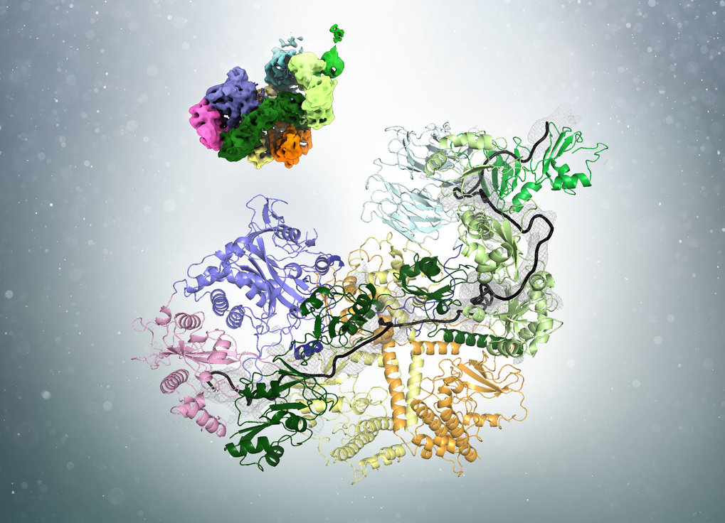
Using cryo-electron microscopy (cryo-EM), the researchers found that PABP forms arches that cover approximately 25 to 30 bases of the poly(A)-tail. Ingmar Schäfer uses an analogy from everyday life to illustrate the process: “Before cutting hair, a hairdresser uses his fingers to physically determine how much of the hair will be left standing.” Similarly, the PABP arches act as a rulers to determine the length of the poly(A)-tail. “But unlike our hair at the hairdresser, the poly(A)-tail is not trimmed with one cut but rather ‘nibbled off’ from the end by the deadenylase enzyme.”

Interestingly, the number of bound PABP alters the affinity of the deadenylase for the poly(A)-tail. In yeast, the most common poly(A)-tail length is around 30 nucleotides. On longer poly(A)-tails, several PABP can be bound and the adenine chain is quickly degraded. Shorter poly(A)-tails approaching the optimal length can only bind one ruler protein. This corresponds with a lower affinity for the deadenylases. “Hence, PABP is not only the tool measuring the poly(A)-tail, but also acts as a brake on the deadenylases when the optimal length is reached”, explains Schäfer. Eventually, when the mRNA is no longer needed, it is further degraded by a different set of deadenylases.

Polyadenylation is a basic principle of cell biology used to control mRNA stability and protein synthesis. “The structure of the poly(A)-tail ‘trimming tool’ will impact research on a broad variety of aspects of cell biology”, puts MPIB Director Elena Conti the study in a larger context. She highlights that the mechanism is highly conserved between yeasts and humans. “Now that we have solved the structure of the deadenylation machinery in yeast, we want to understand how the system works in human cells, where the optimal poly(A)-tail length differs from yeast.”

read more