News

Publication Placeholder

Sahasrabudhe P, Rohrberg J, Biebl MM, Rutz DA, Buchner J.
Mol Cell, 2017, [Epub ahead of print].
doi: 10.1016/j.molcel.2017.08.004.

The Plasticity of the Hsp90 Co-chaperone System.

The Hsp90 system in the eukaryotic cytosol is characterized by a cohort of co-chaperones that bind to Hsp90 and affect its function. Although progress has been made regarding the underlying biochemical mechanisms, how co-chaperones influence Hsp90 client proteins in vivo has remained elusive. By investigating the effect of 12 Hsp90 co-chaperones on the activity of different client proteins in yeast, we find that deletion of co-chaperones can have a neutral or negative effect on client activity but can also lead to more active clients. Only a few co-chaperones are active on all clients studied. Closely related clients and even point mutants can depend on different co-chaperones. These effects are direct because differences in client-co-chaperone interactions can be reconstituted in vitro. Interestingly, some co-chaperones affect client conformation in vivo. Thus, co-chaperones adapt the Hsp90 cycle to the requirements of the client proteins, ensuring optimal activation.


Multiple sclerosis (MS) is the most common inflammatory disease of the central nervous system. It has been suspected for some time that bacteria in the natural intestinal flora may be responsible for triggering the disease in individuals genetically predisposed to it. Together with researchers from the Ludwig Maximilian University of Munich, the Max Planck Institute of Immunobiology and Epigenetics in Freiburg and the Universities of California (San Francisco) and Münster, Hartmut Wekerle and Gurumoorthy Krishnamoorthy from the Max Planck Institutes of Neurobiology and of Biochemistry in Martinsried have, for the first time, shown that intestinal flora from patients with MS can trigger an MS-like illness in an animal model.

n autoimmune diseases such as multiple sclerosis, errant immune cells attack the body's own cells in the brain and spinal cord. Attacks triggered by autoimmune T-cells damage the nerve cells and result in the destruction of the sheath that surrounds these cells. The cells die off, with the result that nerve impulses are no longer transmitted correctly.

Every person has T-cells with the potential to attack their own cells; however, these cells usually remain permanently dormant. In some people, however, the pathogenic potential of these cells is activated, resulting in MS. Scientists believe that activation is caused by a combination of genetic and environmental factors. "More than 200 genes that increase susceptibility to MS have now been identified," explains Hartmut Wekerle, Hertie Professor and Emeritus Director at the Max Planck Institute of Neurobiology, “but for MS to develop, there must be a trigger. To date, most research on triggers has looked at infectious diseases.” A few years ago, the neuroimmunologist, together with his colleagues Kerstin Berer and Gurumoorthy Krishnamoorthy who now leads a Research Group at the MPI of Biochemistry, determined that this trigger is likely to be found in the natural intestinal flora. Together with other colleagues, the researchers showed that intestinal microorganisms were able to activate T-cells in genetically modified autoimmune mice, causing the mice to develop brain lesions similar to those found in MS.

read more


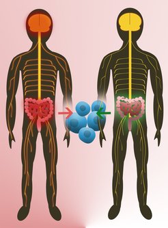
A common feature of neurodegenerative diseases such as Alzheimer’s, Parkinson’s, and Huntington’s is the accumulation of toxic protein deposits in the nerve cells of patients. Once these aggregates appear, they begin to proliferate like weeds. If and how these deposits damage nerve cells and lead to their demise remains largely unexplained. A detailed insight into the three-dimensional structure of the protein aggregates should help researchers to solve this puzzle. Now, using cryo-electron tomography, scientists at the Max Planck Institute of Biochemistry in Martinsried near Munich have succeeded in generating a high-resolution, three-dimensional model of the huntingtin aggregates responsible for Huntington’s disease. The results are published in the journal Cell.

Rampant weed growth – the nightmare of every hobby gardener. Trimming, cropping, cutting. Thorough garden maintenance is required. If this maintenance is neglected, weeds gain the upper hand and suppress the growth of crop and ornamental plants. The same applies to proteins in our bodies: molecular machines, large protein complexes that control vital cellular processes, assume the responsibility of a gardener. These molecular machines ensure that proteins reach their correct conformations and tend to and care for them for the duration of their lifespans.

A matter of the correct form
In order to carry out its function, a protein needs to adopt its correct three-dimensional structure. The building blocks of proteins, the amino acids, are assembled into long chains and folded into a complex form. If the resulting structure is faulty, the defective proteins are broken down in a strictly regulated process. If this does not occur properly, the misfolded proteins may aggregate forming clumps and deposits. Insoluble protein aggregates are toxic for cells. In the brain of patients suffering from neurodegenerative diseases such as Alzheimer’s, Parkinson’s, or Huntington’s, protein aggregates are often found.

If and how exactly these aggregates exert their toxic effects has not yet been explained. This is the question studied by the ToPAG (Toxic Protein AGgregation in neurodegeneration) consortium. A team of researchers in the departments of Wolfgang Baumeister, Ulrich Hartl and Rüdiger Klein has succeeded in decoding a 3D structure of the protein aggregates linked to Huntington’s disease within their intact cellular environment.

read more


Publication Placeholder

Wilhelm L. and Gruber S.
Methods Mol Biol. (2017), 1624, 117-126.
doi: 10.1007/978-1-4939-7098-8_10.


A Chromosome Co-Entrapment Assay to Study Topological Protein-DNA Interactions.

Chromosome organization, DNA replication, and transcription are only some of the processes relying on dynamic and highly regulated protein-DNA interactions. Here, we describe a biochemical assay to study the molecular details of associations between ring-shaped protein complexes and chromosomes in the context of living cells. Any protein complex embracing chromosomal DNA can be enriched by this method, allowing for the underlying loading mechanisms to be investigated.


Brain region mediates pleasure of eating

Providing the body with food is essential for survival. But even when full, we can still take pleasure in eating. Researchers at the Max Planck Institute of Neurobiology in Martinsried and the Friedrich Miescher Institute in Basel have characterized a type of neuron in the amygdala of the mouse brain that is involved in making eating rewarding. When given the choice, mice choose to activate these amygdala neurons. Artificially activating these neurons increases food intake even when the mice are not hungry. The neurobiologists have identified the neuronal circuitry underlying this behavior, raising the possibility that there could be cells with a similar function in the human brain.

The amygdala in the brain plays a key role in emotional responses, decision-making and association of events with emotions like fear or pleasure. In recent years, it has become apparent that this brain region also plays a role in eating behavior. Researchers at the California Institute of Technology have previously shown that activating a certain type of neurons in the amygdala (known as PKC-delta neurons) causes mice to stop eating. “If the mice eat something which has gone bad, for example, activity of these cells causes them to immediately stop eating,” explains Rüdiger Klein, Director at the Max Planck Institute of Neurobiology. “I found this study on ‘anorexia neurons’ in the amygdala fascinating,” says Klein, “so when three doctoral students with very different methodological backgrounds came to me, I proposed them to work on the amygdala project. Their task was to find out whether there are neurons that are involved in positively regulating food consumption.” With this task in mind, the group focused on a different population of amygdala cells named HTR2a neurons.”

Specializing in behavior, electrophysiology and anatomy, the three doctoral students were able to provide insight into HTR2a cell function from a range of angles. “It was a very collaborative project,” recalls Amelia Douglass, one of the three lead authors of the study, which was published in Nature Neuroscience. “We frequently sat down together, went through the results and then built on them, applying new cutting-edge methods in the process.” Using this approach, the young researchers gradually discovered the role of the previously unstudied HTR2a amygdala cells and identified the neural circuitry involved. “Basically we showed that HTR2a cells have a positive effect on food consumption in mice, and that the mice like it when these cells are active,” says Douglass.

read more


Using light alone, scientists from the Max Planck Institute of Neurobiology in Martinsried are now able to reveal pairs or chains of functionally connected neurons under the microscope. The new optogenetic method, named Optobow, allows probing the pathways along which information flows by targeted activation of individual neurons and monitoring the responses of neighboring cells. The shape of the cells and their contact points are also revealed – even in dense tissue in which the thin fibers of thousands of cells are interwoven. With Optobow, it is thus possible to discover individual components of functional circuits in the living brain.

Modern methods provide increasingly detailed insights into the structure and functions of the brain. It is now possible to observe under the microscope when and where neurons are active during a particular task, such as sensory perception or behavior. However, it is still largely impossible to establish whether the active cells are connected to each other and to identify the sequence in which they exchange information. To date, such information could only be obtained, in part and with considerable effort, using electrophysiology and electron microscopy methods.

With electrophysiology, the activity of neighboring cells is measured using very thin, hollow needles, which serve as electrodes. These are inserted into the brain through holes in the skin and the skull of the animal. However, it is almost impossible to record activity from very small, densely clustered or deep-lying neurons, and it is also difficult to trace long connection pathways between neurons. Moreover, impulses from only one, or few cells, can be recorded at a time. With modern electron microscopy processes (connectomics), all neurons and their connections in a fixed brain are recorded, layer by layer, by a scanning electron microscope and then reconstructed on a computer. Although this method provides a fantastically detailed snapshot of the neural wiring pattern, the information about the dynamic transmission of nerve impulses in the living brain is lost. Both of these approaches thus have clear limitations. “We were looking for a way to observe the connections and transmission of information between nerve cells in the active brain without killing or disrupting the brain,” explains Dominique Förster. Motivated by this quest, Förster and his colleagues from Herwig Baier’s Department at the Max Planck Institute of Neurobiology developed the Optobow method.

read more


Understanding brain function generally requires a deep engagement with the matter at least at two separate levels. These are the level of behavioral algorithms on the one hand and the level of neural implementation on the other. These questions can be rephrased as “What exactly is the brain doing?” and “How does it do whatever it is doing?”. Central to this division of labor in the brain sciences is the idea, inspired by David Marr, that the brain implements algorithms, whether for sensory processing, decision making, or motor control, and that these algorithms can be inferred from careful observation of ethologically relevant behaviors. Once a particular algorithm is understood and delineated, we can interrogate its neural implementation by measuring from and manipulating the underlying neural circuits in the context of behavior. Uncovering behavioral algorithms usually does not require modern and cutting edge technologies like optogenetics, wholebrain imaging and genetic editing, it merely requires precise observation, thorough experimental design and, most importantly, rigorous and deep thinking. Such basic scientific qualities are somewhat threatened, and often considered quaint, in the modern era of big data, high throughput and cutting edge technologies. In light of these concerns it comes as a truly pleasant surprise that a study based exclusively on such somewhat antiquated techniques can still find a place in a high impact journal and generate plenty of enthusiasm in the scientific community. The study published recently in Nature by the group of Florian Engert at Harvard in collaboration with the group of Ruben Portugues at the Max Planck Institute of Neurobiology relied entirely on the old-fashioned technique of careful behavioral observations and could have been accomplished in the same form half a century ago. As such it might as well considered “timeless”.

In short, they wanted to know how a larval zebrafish, when placed into a flowing body of water, can detect the presence of the current and then effectively swim against it. More precisely, the group, spearheaded by the team of Pablo Oteiza and Iris Odstrcil, identified the lateral line as one of the main sensory modalities the fish can use to detect that it is drifting with respect to the shore – and more importantly - they could uncover the precise algorithms that translate this sensory input into the appropriate motor commands.

read more


Publication Placeholder

Pünzeler S, Link S, Wagner G, Keilhauer EC, Kronbeck N, Spitzer RM, Leidescher S, Markaki Y, Mentele E, Regnard C, Schneider K, Takahashi D, Kusakabe M, Vardabasso C, Zink LM, Straub T, Bernstein E, Harata M, Leonhardt H, Mann M, Rupp RA, Hake SB.
EMBO J, 2017, doi: 10.15252/embj.201695757. [Epub ahead of print]

Multivalent binding of PWWP2A to H2A.Z regulates mitosis and neural crest differentiation.

Replacement of canonical histones with specialized histone variants promotes altering of chromatin structure and function. The essential histone variant H2A.Z affects various DNA-based processes via poorly understood mechanisms. Here, we determine the comprehensive interactome of H2A.Z and identify PWWP2A as a novel H2A.Z-nucleosome binder. PWWP2A is a functionally uncharacterized, vertebrate-specific protein that binds very tightly to chromatin through a concerted multivalent binding mode. Two internal protein regions mediate H2A.Z-specificity and nucleosome interaction, whereas the PWWP domain exhibits direct DNA binding. Genome-wide mapping reveals that PWWP2A binds selectively to H2A.Z-containing nucleosomes with strong preference for promoters of highly transcribed genes. In human cells, its depletion affects gene expression and impairs proliferation via a mitotic delay. While PWWP2A does not influence H2A.Z occupancy, the C-terminal tail of H2A.Z is one important mediator to recruit PWWP2A to chromatin. Knockdown of PWWP2A in Xenopus results in severe cranial facial defects, arising from neural crest cell differentiation and migration problems. Thus, PWWP2A is a novel H2A.Z-specific multivalent chromatin binder providing a surprising link between H2A.Z, chromosome segregation, and organ development.


Red, green and blue: normally, no more than three different colours can be detected simultaneously in fluorescence microscopy. Thanks to recent RGB nanotechnology, similar to that used in computer monitors, it is now possible to generate 124 virtual colours under the microscope. The three primary colours are arranged in various mixing ratios on a special DNA lattice. This creates individual colour pixels under the microscope. The new method was developed by scientists at the Max Planck Institute of Biochemistry, Ludwig Maximilian University of Munich, Germany, and the Wyss Institute for Biologically Inspired Engineering at Harvard University in the US. The team’s work was published in the journal Science Advances

Biomedical research has made tremendous strides in recent decades. Using the latest microscopes, scientists are analyzing the function and interactions of molecules in cells with ever greater detail. Now, researchers are looking for methods to image multiple molecules simultaneously.

RGB nanotechnology
A team of scientists from Germany and the US headed by Ralf Jungmann and Peng Yin has now developed substances known as metafluorophors. “The technology can be compared to that of an RGB monitor,” explains Jungmann, Leader of the Molecular Imaging and Bionanotechnology Research Group. To display a wide range of colours on a screen, each colour is mixed from the three primary colours: red, green and blue. “We’ve transferred this approach to the nanometre scale. Instead of a single fluorescence dye molecule, multiple fluorescent molecules are applied to a carrier material, which serves as a kind of experimental board. Depending on the proportion of the three primary colours, they appear in different colours under the microscope, comparable to nanometre-scale colour pixels on a computer screen.“

read more


Publication Placeholder

Jonak K, Zagoriy I, Oz T, Graf P, Rojas J, Mengoli V, Zachariae W.
Cell Cycle, 2017, [Epub ahead of print].

APC/C-Cdc20 mediates deprotection of centromeric cohesin at meiosis II in yeast.

Cells undergoing meiosis produce haploid gametes through one round of DNA replication followed by 2 rounds of chromosome segregation. This requires that cohesin complexes, which establish sister chromatid cohesion during S phase, are removed in a stepwise manner. At meiosis I, the separase protease triggers the segregation of homologous chromosomes by cleaving cohesin's Rec8 subunit on chromosome arms. Cohesin persists at centromeres because the PP2A phosphatase, recruited by the shugoshin protein, dephosphorylates Rec8 and thereby protects it from cleavage. While chromatids disjoin upon cleavage of centromeric Rec8 at meiosis II, it was unclear how and when centromeric Rec8 is liberated from its protector PP2A. One proposal is that bipolar spindle forces separate PP2A from Rec8 as cells enter metaphase II. We show here that sister centromere biorientation is not sufficient to "deprotect" Rec8 at meiosis II in yeast. Instead, our data suggest that the ubiquitin-ligase APC/CCdc20 removes PP2A from centromeres by targeting for degradation the shugoshin Sgo1 and the kinase Mps1. This implies that Rec8 remains protected until entry into anaphase II when it is phosphorylated concurrently with the activation of separase. Here, we provide further support for this model and speculate on its relevance to mammalian oocytes.