News

Publication Placeholder

Kremer, M.C., Jung, C., Batelli, S., Rubin, G.M., and Gaul, U..
Glia, 2017, [Epub ahead of print].

The glia of the adult Drosophila nervous system.

Glia play crucial roles in the development and homeostasis of the nervous system. While the GLIA in the Drosophila embryo have been well characterized, their study in the adult nervous system has been limited. Here, we present a detailed description of the glia in the adult nervous system, based on the analysis of some 500 glial drivers we identified within a collection of synthetic GAL4 lines. We find that glia make up ∼10% of the cells in the nervous system and envelop all compartments of neurons (soma, dendrites, axons) as well as the nervous system as a whole. Our morphological analysis suggests a set of simple rules governing the morphogenesis of glia and their interactions with other cells. All glial subtypes minimize contact with their glial neighbors but maximize their contact with neurons and adapt their macromorphology and micromorphology to the neuronal entities they envelop. Finally, glial cells show no obvious spatial organization or registration with neuronal entities. Our detailed description of all glial subtypes and their regional specializations, together with the powerful genetic toolkit we provide, will facilitate the functional analysis of glia in the mature nervous system. GLIA 2017.

 

Publication Placeholder

Matscheko, N., Mayrhofer, P., and Wollert, T.
Autophagy, 2017, [Epub ahead of print].

Passing membranes to autophagy: Unconventional membrane tethering by Atg17.

Macroautophagy delivers cytoplasmic material to lysosomal/vacuolar compartments for degradation. Conserved multi-subunit complexes, composed of autophagy related (Atg) proteins, initiate the formation of membrane precursors, termed phagophores. Under physiological conditions these cup- shaped structures can capture cytoplasmic material highly selectively. Starvation or cytotoxic stresses, however, initiate the formation of much larger phagophores to enclose cytoplasm nonselectively. The biogenesis of nonselective autophagosomes is initiated by the hierarchical assembly of the Atg1 kinase complex and the recruitment of Atg9 vesicles at the phagophore assembly site (PAS). In this punctum we summarize our recent findings regarding tethering of Atg9 vesicles by the Atg1 kinase complex. We discuss membrane tethering by and activation of its central subunit Atg17 in the context of other canonical membrane tethering factors. Our results show that Atg17 suffices to bind and tether Atg9 vesicles. The Atg31-Atg29 subcomplex inhibits Atg17 activity, and activation of Atg17 depends on the formation of the Atg1 kinase complex that involves recruiting Atg1-Atg13. Our studies lead to a model of unconventional membrane tethering in autophagy.


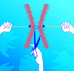
Once together, never apart – isn’t that how the saying goes? Not so in meiosis, the special type of cell division in which gametes, sperm and egg cells are formed. At the start of meiosis the ring-shaped protein complex, referred to as cohesin, is the string that ties the chromosome strands together. The chromosomes are where the blueprint for the body is stored. If each egg cell and each sperm is to come out of meiosis with only one set of chromosomes, these strings need to be cut up in a precise pattern. Scientists from the Max Planck Institute of Biochemistry have demonstrated in baker’s yeast how a kinase enzyme, which is also present in the human body, controls the cleavage of the cohesin rings and coordinates it with the exit from meiosis and gamete formation. It is a mechanism, which could explain how chromosome segregation is regulated, or goes wrong, in human sperm and egg cells.

Why is it that children look like their parents? Most of the cells in our body are diploid, which means that they have two copies of each chromosome – one from the mother and one from the father. Only gametes, in other words egg and sperm cells, contain a single copy. So the cell has to halve its double set of chromosomes in order to be able to make haploid gametes. This happens in a special type of cell division called meiosis. However, meiosis is surprisingly complicated. Instead of just dividing up the maternal and paternal chromosomes between two daughter cells, the chromosomes are first duplicated so that they each consist of two strands, or chromatids. Duplicated paternal and maternal chromosomes then align and the chromosome arms are spliced together. This produces new chromosomes out of four chromatids held together by ring-shaped protein complexes, the cohesins, which act like strings. Two divisions of the nucleus are needed in order to split these chromosomes up into their chromatids again in processes referred to as the first and the second meiotic divisions. In the first division, the cohesin rings on the chromosome arms are cut up by the enzyme separase, which acts like a molecular pair of scissors. This results in X-shaped chromosomes consisting of two chromatids held together by cohesin rings only in their centre, known as the centromer. The second meiotic division is when the separase scissors ultimately cut the cohesin rings at the centromer. Four haploid gamete nuclei are produced, each of which contains one chromatid from each chromosome, in other words a blueprint for the body. If the egg is fertilized, the genetic material from the mother and the father combines and a new diploid chromosome set is produced, or to put it simply: a baby with daddy’s nose and mummy’s eyes.

read more


Publication Placeholder

Princz, L.N., Wild, P., Bittmann, J., Aguado, F.J., Blanco, M.G., Matos, J., and Pfander, B.
EMBO J, 2017, [Epub ahead of print].

Dbf4-dependent kinase and the Rtt107 scaffold promote Mus81-Mms4 resolvase activation during mitosis.

DNA repair by homologous recombination is under stringent cell cycle control. This includes the last step of the reaction, disentanglement of DNA joint molecules (JMs). Previous work has established that JM resolving nucleases are activated specifically at the onset of mitosis. In case of budding yeast Mus81-Mms4, this cell cycle stage-specific activation is known to depend on phosphorylation by CDK and Cdc5 kinases. Here, we show that a third cell cycle kinase, Cdc7-Dbf4 (DDK), targets Mus81-Mms4 in conjunction with Cdc5-both kinases bind to as well as phosphorylate Mus81-Mms4 in an interdependent manner. Moreover, DDK-mediated phosphorylation of Mms4 is strictly required for Mus81 activation in mitosis, establishing DDK as a novel regulator of homologous recombination. The scaffold protein Rtt107, which binds the Mus81-Mms4 complex, interacts with Cdc7 and thereby targets DDK and Cdc5 to the complex enabling full Mus81 activation. Therefore, Mus81 activation in mitosis involves at least three cell cycle kinases, CDK, Cdc5 and DDK Furthermore, tethering of the kinases in a stable complex with Mus81 is critical for efficient JM resolution.


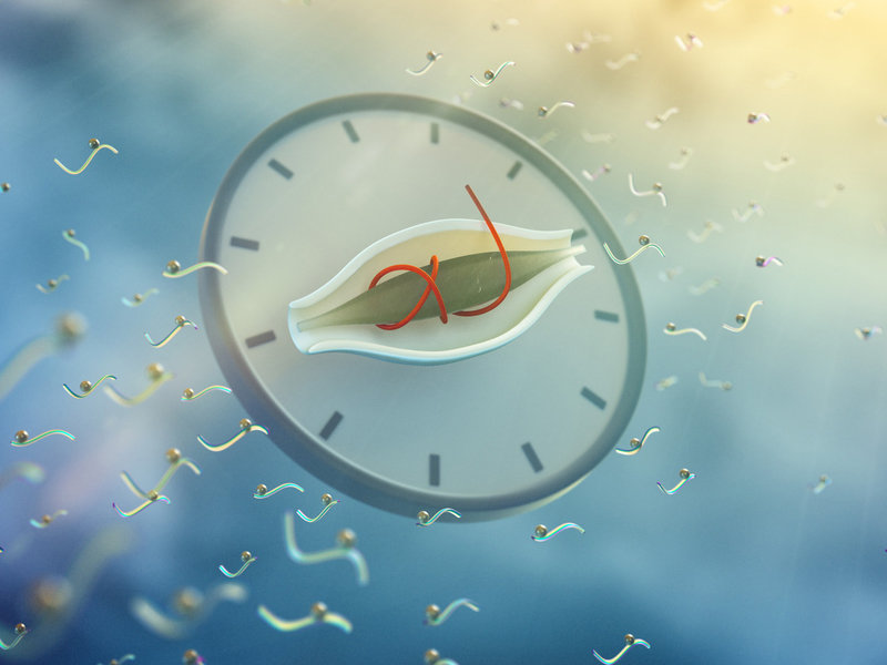
Circadian is the latin meaning for “about a day”. Circadian clocks have evolved to adapt our lives to the daily environmental changes on earth: light and warmth during the day and darkness and cold at night. Scientists at the Max-Planck-Institute of Biochemistry in Martinsried discovered with the help of the mass spectrometry, that more than 25 percent of the molecular protein switches in mouse liver cells change in a daily manner. These rhythmic switches are binding sites for phosphate molecules, that regulate the function of proteins, and thereby the daily metabolic processes in the organ. The study was published in the journal Cell Metabolism.

read more


Publication Placeholder

Bantele, S.C., Ferreira, P., Gritenaite, D., Boos, D., and Pfander, B.
Elife, 2017, 6.

Targeting of the Fun30 nucleosome remodeller by the Dpb11 scaffold facilitates cell cycle-regulated DNA end resection.

DNA double strand breaks (DSBs) can be repaired by either recombination-based or direct ligation-based mechanisms. Pathway choice is made at the level of DNA end resection, a nucleolytic processing step, which primes DSBs for repair by recombination. Resection is thus under cell cycle control, but additionally regulated by chromatin and nucleosome remodellers. Here we show that both layers of control converge in the regulation of resection by the evolutionarily conserved Fun30/SMARCAD1 remodeller. Yeast Fun30 and human SMARCAD1 are cell cycle-regulated by interaction with the DSB-localized scaffold proteins Dpb11 and TOPBP1, respectively. In yeast this protein assembly additionally comprises the 9-1-1 damage sensor, is involved in localizing Fun30 to damaged chromatin and thus is required for efficient long-range resection of DSBs. Notably, artificial targeting of Fun30 to DSBs is sufficient to bypass the cell cycle regulation of long-range resection, indicating that chromatin remodelling during resection is underlying DSB repair pathway choice.


Publication Placeholder

Thanisch, K., Song, C., Engelkamp, D., Koch, J., Wang, A., Hallberg, E., Foisner, R., Leonhardt, H., Stewart, C.L., Joffe, B., and Solovei, I.
Differentiation, 2017, 94, 58-70.

Nuclear envelope localization of LEMD2 is developmentally dynamic and lamin A/C dependent yet insufficient for heterochromatin tethering.

Peripheral heterochromatin in mammalian nuclei is tethered to the nuclear envelope by at least two mechanisms here referred to as the A- and B-tethers. The A-tether includes lamins A/C and additional unknown components presumably INM protein(s) interacting with both lamins A/C and chromatin. The B-tether includes the inner nuclear membrane (INM) protein Lamin B-receptor, which binds B-type lamins and chromatin. Generally, at least one of the tethers is always present in the nuclear envelope of mammalian cells. Deletion of both causes the loss of peripheral heterochromatin and consequently inversion of the entire nuclear architecture, with this occurring naturally in rod photoreceptors of nocturnal mammals. The tethers are differentially utilized during development, regulate gene expression in opposite manners, and play an important role during cell differentiation. Here we aimed to identify the unknown chromatin binding component(s) of the A-tether. We analyzed 10 mouse tissues by immunostaining with antibodies against 7 INM proteins and found that every cell type has specific, although differentially and developmentally regulated, sets of these proteins. In particular, we found that INM protein LEMD2 is concomitantly expressed with A-type lamins in various cell types but is lacking in inverted nuclei of rod cells. Truncation or deletion of Lmna resulted in the downregulation and mislocalization of LEMD2, suggesting that the two proteins interact and pointing at LEMD2 as a potential chromatin binding mediator of the A-tether. Using nuclei of mouse rods as an experimental model lacking peripheral heterochromatin, we expressed a LEMD2 transgene alone or in combination with lamin C in these cells and observed no restoration of peripheral heterochromatin in either case. We conclude that in contrary to the B-tether, the A-tether has a more intricate composition and consists of multiple components that presumably vary, at differing degrees of redundancy, between cell types and differentiation stages.


Publication Placeholder

Hamzeiy, H., and Cox, J.
Curr Opin Biotechnol, 2016, 43, 141-146.

What computational non-targeted mass spectrometry-based metabolomics can gain from shotgun proteomics.

Computational workflows for mass spectrometry-based shotgun proteomics and untargeted metabolomics share many steps. Despite the similarities, untargeted metabolomics is lagging behind in terms of reliable fully automated quantitative data analysis. We argue that metabolomics will strongly benefit from the adaptation of successful automated proteomics workflows to metabolomics. MaxQuant is a popular platform for proteomics data analysis and is widely considered to be superior in achieving high precursor mass accuracies through advanced nonlinear recalibration, usually leading to five to ten-fold better accuracy in complex LC-MS/MS runs. This translates to a sharp decrease in the number of peptide candidates per measured feature, thereby strongly improving the coverage of identified peptides. We argue that similar strategies can be applied to untargeted metabolomics, leading to equivalent improvements in metabolite identification.


Publication Placeholder

Geyer, P.E., Wewer Albrechtsen, N.J., Tyanova, S., Grassl, N., Iepsen, E.W., Lundgren, J., Madsbad, S., Holst, J.J., Torekov, S.S., and Mann, M.
Mol Syst Biol, 2016, 12, 901.

Proteomics reveals the effects of sustained weight loss on the human plasma proteome.

Sustained weight loss is a preferred intervention in a wide range of metabolic conditions, but the effects on an individual's health state remain ill-defined. Here, we investigate the plasma proteomes of a cohort of 43 obese individuals that had undergone 8 weeks of 12% body weight loss followed by a year of weight maintenance. Using mass spectrometry-based plasma proteome profiling, we measured 1,294 plasma proteomes. Longitudinal monitoring of the cohort revealed individual-specific protein levels with wide-ranging effects of losing weight on the plasma proteome reflected in 93 significantly affected proteins. The adipocyte-secreted SERPINF1 and apolipoprotein APOF1 were most significantly regulated with fold changes of -16% and +37%, respectively (P < 10-13), and the entire apolipoprotein family showed characteristic differential regulation. Clinical laboratory parameters are reflected in the plasma proteome, and eight plasma proteins correlated better with insulin resistance than the known marker adiponectin. Nearly all study participants benefited from weight loss regarding a ten-protein inflammation panel defined from the proteomics data. We conclude that plasma proteome profiling broadly evaluates and monitors intervention in metabolic diseases.


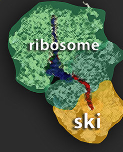
Although the terms “cryo-EM” and “SKI complex” evoke images of ice and snow, they actually relate to structural biology. Scientists at the Max Planck Institute of Biochemistry and the Gene Center of the University of Munich (LMU) have now shown that the cellular protein factory and the SKI protein complex are in direct contact. The SKI complex is part of a molecular shredder that breaks down mRNA, the construction manual for proteins, into its individual components. To conduct their analysis, the researchers used cryo-electron microscopy, a technique that involves flash-freezing protein complexes to allow even the tiniest details of their structure to be studied in their natural state.

Ribosomes are the molecular protein factories of cells. Following a construction manual – the messenger RNA, or mRNA – they assemble protein building blocks to form chains. These chains are later folded into tiny molecular machines – the proteins. Proteins then perform a variety of tasks in the cells. Roland Beckmann and his team at the Gene Center of the University of Munich (LMU) specialize in investigating the structure of the ribosomes using cryo-electron microscopy. Elena Conti’s group “Structural Cell Biology” at the Max Planck Institute of Biochemistry in Martinsried has been studying the structure and function of the exosome, a shredder for mRNA molecules, for many years. When the protein construction manual is no longer needed, or when it contains an error, the exosome breaks down the mRNA into its basic components and recycles it.

In a joint project between these two groups, scientists at the two institutions have now shown that the SKI protein complex, which serves as the motor for the exosome in breaking down mRNA, is in direct contact with the ribosomes.

read more