
News

Publication Placeholder

Choi J, Bachmann AL, Tauscher K, Benda C, Fierz B, Müller J.
Nat Struct Mol Biol, 2017, [Epub ahead of print].
doi: 10.1038/nsmb.3488

DNA binding by PHF1 prolongs PRC2 residence time on chromatin and thereby promotes H3K27 methylation.

Polycomb repressive complex 2 (PRC2) trimethylates histone H3 at lysine 27 to mark genes for repression. We measured the dynamics of PRC2 binding on recombinant chromatin and free DNA at the single-molecule level using total internal reflection fluorescence (TIRF) microscopy. PRC2 preferentially binds free DNA with multisecond residence time and midnanomolar affinity. PHF1, a PRC2 accessory protein of the Polycomblike family, extends PRC2 residence time on DNA and chromatin. Crystallographic and functional studies reveal that Polycomblike proteins contain a winged-helix domain that binds DNA in a sequence-nonspecific fashion. DNA binding by this winged-helix domain accounts for the prolonged residence time of PHF1-PRC2 on chromatin and makes it a more efficient H3K27 methyltranferase than PRC2 alone. Together, these studies establish that interactions with DNA provide the predominant binding affinity of PRC2 for chromatin. Moreover, they reveal the molecular basis for how Polycomblike proteins stabilize PRC2 on chromatin and stimulate its activity.

 

Publication Placeholder

Karg E, Smets M, Ryan J, Forné I, Qin W, Mulholland CB, Kalideris G, Imhof A, Bultmann S, Leonhardt H.
J Mol Biol, 2017, [Epub ahead of print].
doi: 10.1016/j.jmb.2017.10.014

Ubiquitome analysis reveals PCNA-associated factor 15 (PAF15) as a specific ubiquitination target of UHRF1 in embryonic stem cells.

Ubiquitination is a multifunctional posttranslational modification controlling the activity, subcellular localization and stability of proteins. The E3 ubiquitin ligase UHRF1 is an essential epigenetic factor that recognizes repressive histone marks as well as hemi-methylated DNA and recruits DNMT1. To explore enzymatic functions of UHRF1 beyond epigenetic regulation we conducted a comprehensive screen in mouse embryonic stem cells to identify novel ubiquitination targets of UHRF1 and its paralogue UHRF2. We found differentially ubiquitinated peptides associated with a variety of biological processes such as transcriptional regulation and DNA damage response. Most prominently, we identified PCNA-associated factor 15 (PAF15, also known as Pclaf, Ns5atp9, KIAA0101 and OEATC-1) as a specific ubiquitination target of UHRF1. Although the function of PAF15 ubiquitination in translesion DNA synthesis (TLS) is well characterized, the respective E3 ligase had been unknown. We could show that UHRF1 ubiquitinates PAF15 at Lys 15 and Lys 24 and promotes its binding to PCNA during late S-phase. In summary, we identified novel ubiquitination targets that link UHRF1 to transcriptional regulation and DNA damage response.


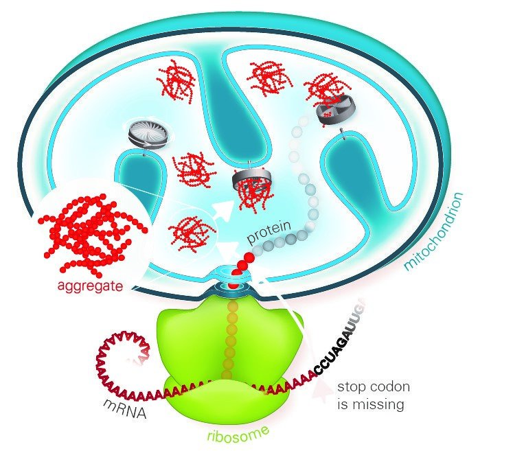
A common feature of neurodegenerative diseases such as Alzheimer's, Parkinson's or Huntington's disease are deposits of aggregated proteins in the patient's cells that cause damage to cellular functions. Scientists at the Max Planck Institute of Biochemistry and Ludwig-Maximilians-Universität in Munich report that, even in normal cells, aberrant aggregation-prone proteins are continually produced due to partial failure of the respiratory system. Unless they are removed by degradation, aggregates accumulate preferentially in the mitochondria, the cellular power plants, ultimately blocking energy production. In order to get rid of these toxic aggregates, cells have developed an elaborate protein quality control system.

Misfolded proteins made from defective blueprints are often sticky and clump together. Accumulation of such faulty proteins is known to contribute to the progression of several diseases. Therefore, cells have internal quality control mechanisms that detect and rapidly destroy faulty proteins. Proteins are produced by ribosomes, and misfolding can occur if they stall while decoding a damaged template. If the necessary ribosome-associated quality control machinery (RQC) does not function properly, defective proteins accumulate and form toxic aggregates in the cytoplasm of the cells. A previous study reported that this aggregation mechanism is mediated by so-called CAT-tails – C-terminal alanine-threonine sequences that are added to the defective proteins. So far, studies have focused on how the RQC recognizes and clears blocked ribosomes in the cytosol. The collaborating groups at the Max Planck Institute of Biochemistry and the university have now investigated the clearance of ribosome-blocked proteins destined for the mitochondria.

read more


Publication Placeholder

Geyer PE, Holdt LM, Teupser D, Mann M.
Mol Syst Biol 2017, 13, 942.
doi: 10.15252/msb.20156297

Revisiting biomarker discovery by plasma proteomics.

Clinical analysis of blood is the most widespread diagnostic procedure in medicine, and blood biomarkers are used to categorize patients and to support treatment decisions. However, existing biomarkers are far from comprehensive and often lack specificity and new ones are being developed at a very slow rate. As described in this review, mass spectrometry (MS)-based proteomics has become a powerful technology in biological research and it is now poised to allow the characterization of the plasma proteome in great depth. Previous "triangular strategies" aimed at discovering single biomarker candidates in small cohorts, followed by classical immunoassays in much larger validation cohorts. We propose a "rectangular" plasma proteome profiling strategy, in which the proteome patterns of large cohorts are correlated with their phenotypes in health and disease. Translating such concepts into clinical practice will require restructuring several aspects of diagnostic decision-making, and we discuss some first steps in this direction.


Publication Placeholder

Pasalic D, Weber B, Giannone C, Anelli T, Müller R, Fagioli C, Felkl M, John C, Mossuto MF, Becker CFW, Sitia R, Buchner J.
Proc Natl Acad Sci USA 2017, [Epub ahead of print].
doi: 10.1073/pnas.1701797114

A peptide extension dictates IgM assembly.

Professional secretory cells can produce large amounts of high-quality complex molecules, including IgM antibodies. Owing to their multivalency, polymeric IgM antibodies provide an efficient first-line of defense against pathogens. To decipher the mechanisms of IgM assembly, we investigated its biosynthesis in living cells and faithfully reconstituted the underlying processes in vitro. We find that a conserved peptide extension at the C-terminal end of the IgM heavy (Ig-μ) chains, termed the tailpiece, is necessary and sufficient to establish the correct geometry. Alanine scanning revealed that hydrophobic amino acids in the first half of the tailpiece contain essential information for generating the correct topology. Assembly is triggered by the formation of a disulfide bond linking two tailpieces. This induces conformational changes in the tailpiece and the adjacent domain, which drive further polymerization. Thus, the biogenesis of large and topologically challenging IgM complexes is dictated by a local conformational switch in a peptide extension.


Publication Placeholder

Li L, Lingaraju M, Basquin C, Basquin J, Conti E
RNA 2017, 23, 1028-1034.
doi: 10.1261/rna.061200.117

Structure of a SMG8-SMG9 complex identifies a G-domain heterodimer in the NMD effector proteins.

Nonsense-mediated mRNA decay (NMD) is a eukaryotic mRNA degradation pathway involved in surveillance and post-transcriptional regulation, and executed by the concerted action of several trans-acting factors. The SMG1 kinase is an essential NMD factor in metazoans and is associated with two recently identified and yet poorly characterized proteins, SMG8 and SMG9. We determined the 2.5 Å resolution crystal structure of a SMG8-SMG9 core complex from C. elegans We found that SMG8-SMG9 is a G-domain heterodimer with architectural similarities to the dynamin-like family of GTPases such as Atlastin and GBP1. The SMG8-SMG9 heterodimer forms in the absence of nucleotides, with interactions conserved from worms to humans. Nucleotide binding occurs at the G domain of SMG9 but not of SMG8. Fitting the GDP-bound SMG8-SMG9 structure in EM densities of the human SMG1-SMG8-SMG9 complex raises the possibility that the nucleotide site of SMG9 faces SMG1 and could impact the kinase conformation and/or regulation.


Publication Placeholder

Varga J & Greten FR
Nat Cell Biol, 2017, [Epub ahead of print].
doi: 10.1038/ncb3611

Cell plasticity in epithelial homeostasis and tumorigenesis

The adult organism is characterized by remarkable plasticity, which enables efficient regeneration and restoration of homeostasis after damage. When aberrantly activated, this plasticity contributes to tumour initiation and progression. Here we review recent advances in this field with a focus on cell fate changes and the epithelial-mesenchymal transition-two distinct, yet closely related, forms of plasticity with fundamental roles in homeostasis and cancer.


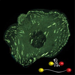
Proteins are often considered as molecular machines. To understand how they work, it is not enough to visualize the involved proteins under the microscope. Wherever machines are at work mechanical forces occur, which in turn influence biological processes. These extremely small intracellular forces can be measured with the help of molecular force sensors. Now researchers at the Max Planck Institute of Biochemistry in Martinsried have developed molecular probes that can measure forces across multiple proteins with high resolution in cells. The results of their work were published in the journal Nature Methods.

When proteins pull on each other, forces in the piconewton range are generated. Cells can detect such mechanical information and modulate their response depending on the nature of the signal. Adhesion proteins on the surface of cells, for instance, recognize how rigid their environment is to adjust the protein composition of the cell accordingly. To measure such tiny forces, the group of Molecular Mechanotransduction at the Max Planck Institute is developing molecular force sensors. “These small measuring instruments work along the lines of a spring scale,” says Carsten Grashoff, head of the research group.

The innovative probes consist of two fluorescent molecules that are connected by a sort of molecular spring. When a force of just a few piconewton acts on the molecule, the spring stretches, and this change can be detected using a special microscopic method. “We’re now able to measure the mechanics of several molecules simultaneously,” Carsten Grashoff explains. In contrast to previous experiments, the scientists are not only able to determine which proteins, but also how many of them are under force at any given moment.

read more


Publication Placeholder

di Cicco G, Bantele SCS, Reusswig KU, Pfander B. (IMPRS-LS students are in bold)
Sci Rep 7, 11650.
doi: 10.1038/s41598-017-11937-z

A cell cycle-independent mode of the Rad9-Dpb11 interaction is induced by DNA damage.

Budding yeast Rad9, like its orthologs, controls two aspects of the cellular response to DNA double strand breaks (DSBs) - signalling of the DNA damage checkpoint and DNA end resection. Rad9 binds to damaged chromatin via modified nucleosomes independently of the cell cycle phase. Additionally, Rad9 engages in a cell cycle-regulated interaction with Dpb11 and the 9-1-1 clamp, generating a second pathway that recruits Rad9 to DNA damage sites. Binding to Dpb11 depends on specific S/TP phosphorylation sites of Rad9, which are modified by cyclin-dependent kinase (CDK). Here, we show that these sites additionally become phosphorylated upon DNA damage. We define the requirements for DNA damage-induced S/TP phosphorylation of Rad9 and show that it is independent of the cell cycle or CDK activity but requires prior recruitment of Rad9 to damaged chromatin, indicating that it is catalysed by a chromatin-bound kinase. The checkpoint kinases Mec1 and Tel1 are required for Rad9 S/TP phosphorylation, but their influence is likely indirect and involves phosphorylation of Rad9 at S/TQ sites. Notably, DNA damage-induced S/TP phosphorylation triggers Dpb11 binding to Rad9, but the DNA damage-induced Rad9-Dpb11 interaction is dispensable for recruitment to DNA damage sites, indicating that the Rad9-Dpb11 interaction functions beyond Rad9 recruitment.


Publication Placeholder

Andreeva L, Hiller B, Kostrewa D, Lässig C, de Oliveira Mann CC, Jan Drexler D, Maiser A, Gaidt M, Leonhardt H, Hornung V, Hopfner KP.
Nature, 2017,  [Epub ahead of print].
doi: 10.1038/nature23890.

cGAS senses long and HMGB/TFAM-bound U-turn DNA by forming protein-DNA ladders.

Cytosolic DNA arising from intracellular pathogens triggers a powerful innate immune response. It is sensed by cyclic GMP-AMP synthase (cGAS), which elicits the production of type I interferons by generating the second messenger 2'3'-cyclic-GMP-AMP (cGAMP). Endogenous nuclear or mitochondrial DNA can also be sensed by cGAS under certain conditions, resulting in sterile inflammation. The cGAS dimer binds two DNA ligands shorter than 20 base pairs side-by-side, but 20-base-pair DNA fails to activate cGAS in vivo and is a poor activator in vitro. Here we show that cGAS is activated in a strongly DNA length-dependent manner both in vitro and in human cells. We also show that cGAS dimers form ladder-like networks with DNA, leading to cooperative sensing of DNA length: assembly of the pioneering cGAS dimer between two DNA molecules is ineffective; but, once formed, it prearranges the flanking DNA to promote binding of subsequent cGAS dimers. Remarkably, bacterial and mitochondrial nucleoid proteins HU and mitochondrial transcription factor A (TFAM), as well as high-mobility group box 1 protein (HMGB1), can strongly stimulate long DNA sensing by cGAS. U-turns and bends in DNA induced by these proteins pre-structure DNA to nucleate cGAS dimers. Our results suggest a nucleation-cooperativity-based mechanism for sensitive detection of mitochondrial DNA and pathogen genomes, and identify HMGB/TFAM proteins as DNA-structuring host factors. They provide an explanation for the peculiar cGAS dimer structure and suggest that cGAS preferentially binds incomplete nucleoid-like structures or bent DNA.